
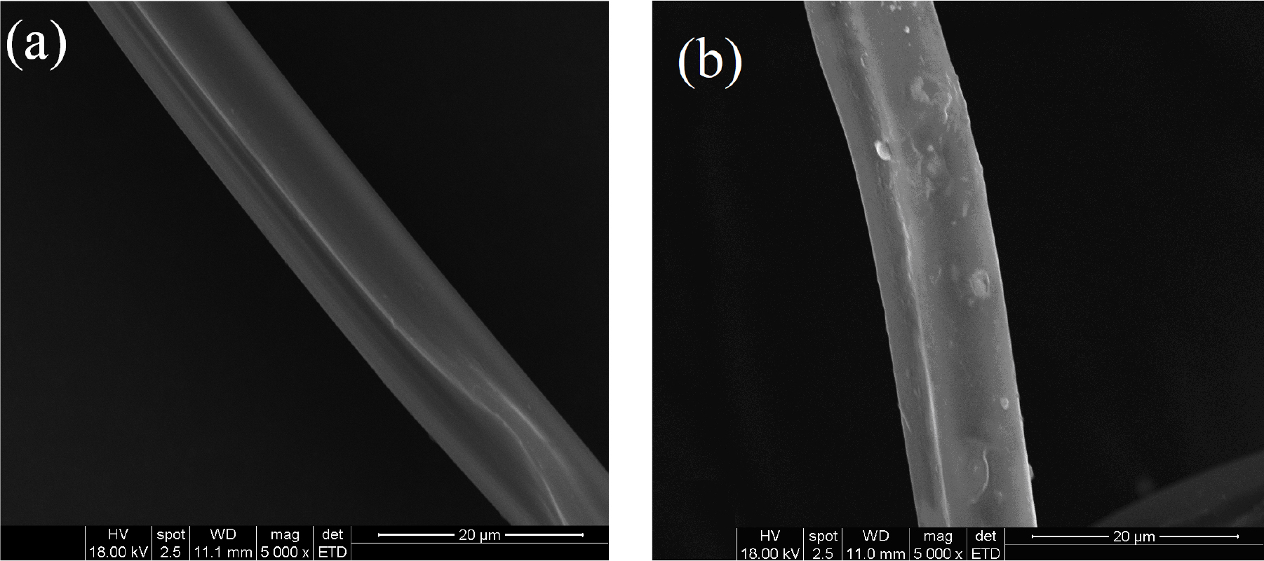
Fig. 1.
Longitudinal surface photomicrographs of (a) untreated silk fiber and (b) modified silk fiber treated with NMA-HTCC (×5000)
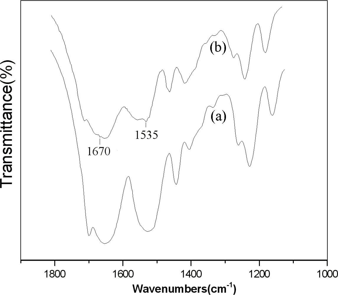
Fig. 2.
FT-IR spectroscopy of (a) B. mori silk fiber and (b) modified silk fiber treated with NMA-HTCC
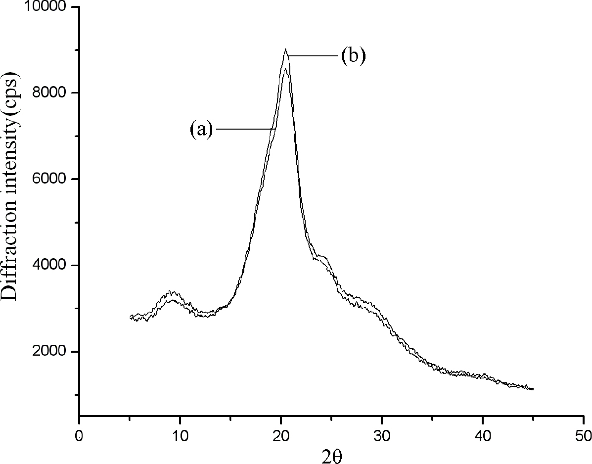
Fig. 3.
XRD curve peak fitting of (a) Bombyx mori silk fiber, (b) NMA-HTCC modified silk fiber
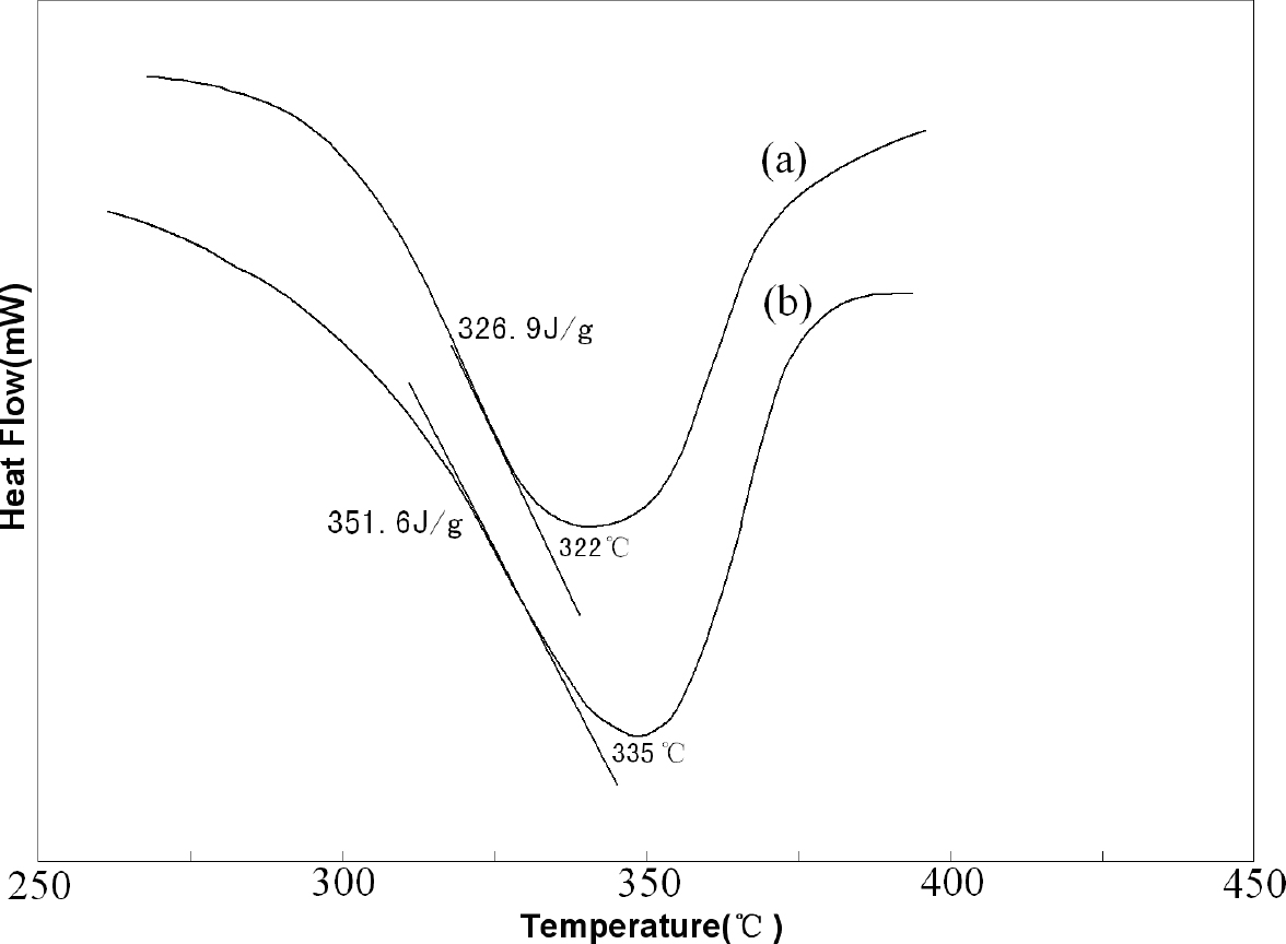
Fig. 4.
DSC diagram of (a) Bombyx mori silk and (b) modified silk treated with NMA-HTCC
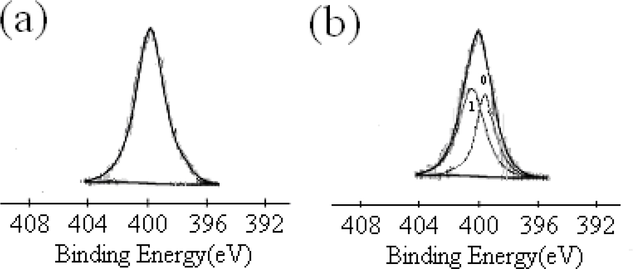
Fig. 5.
XPS spectra of N1s peaks for silk fibers: (a) before treatment, (b) after treatment with NMA-HTCC
Table 1.
Composition analysis of N(1s)
| Parameter of peak | Before treatment | After treatment | |
|---|---|---|---|
| 0 peak | 1 peak | ||
| Binding energy/eV | 399.70 | 399.50 | 400.50 |
| Half peak width/eV | 2.13 | 1.89 | 1.99 |
| Peak area | 251.67 | 68.16 | 79.68 |
Table 2.
Elements on the surface of silk fiber
| Element composition | Surface element content of silk fiber (%) | Change rate (%) | |
|---|---|---|---|
| Before treatment | After treatment | ||
| C | 83.44 | 86.19 | + 3.30 |
| O | 9.42 | 8.79 | -6.69 |
| N | 5.20 | 2.19 | -57.88 |
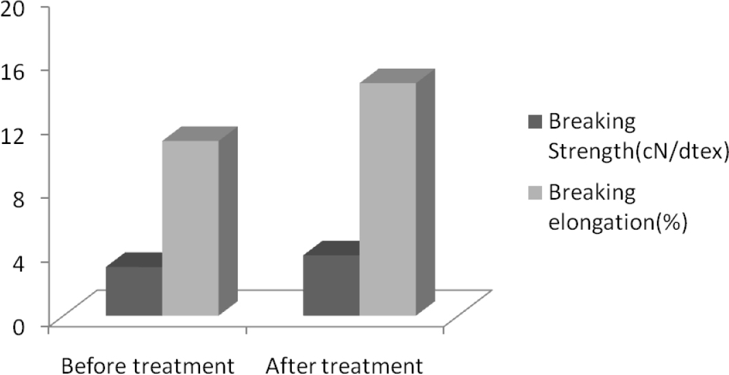
Fig. 6.
Breaking strength and breaking elongation of silk fibers
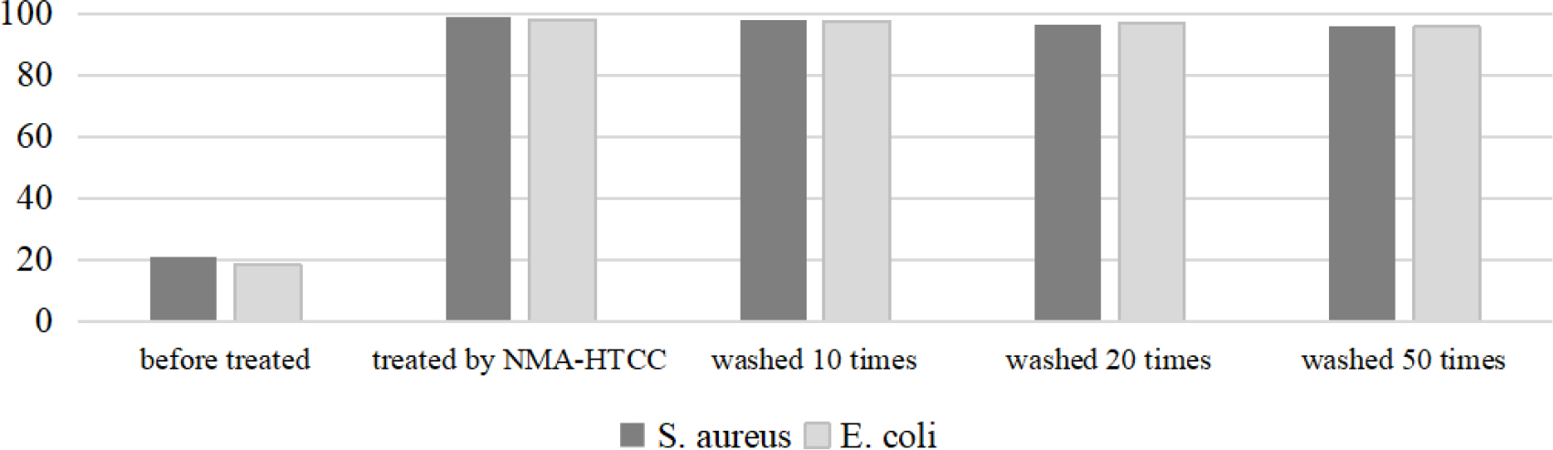
Fig. 7.
Antibacterial activities of silk fibers