
Table 1.
Main reagents
| Laboratory reagent | Molecular formula | Molecular weight | Specifications | Character | Manufacturer |
|---|---|---|---|---|---|
| Pyrrole | C4H5N | 67.09 | Chemically pure | Yellowish transparent oily liquid | Shanghai Kefeng Industrial Co., Ltd |
| Ammonium persulfate | (NH4)2S2O8 | 228.18 | Analytically pure | White powder | Tianjin Wind Ship Chemical Technology Co., Ltd |
| p-toluene sulfonic acid | C7H8O3S | 172.20 | Analytically pure | White crystalline | Tianjin Guangfu Fine Chemicals Institute |
Table 2.
Main experimental instruments
| Instrument | Specifications | Manufacturer |
|---|---|---|
| Intelligent digital display constant temperature water bath | HH-S4 | Gongyi City Yuhua Instrument Co., Ltd, China |
| Desktop digital multimeter | 34410A | Agilent Technologies, USA |
| Field emission scanning electron microscope | S-4800 | Hitachi Ltd, Japan |
Table 3.
Process parameters
| Doping agent (mol/L) | Mole ratio of oxidant and pyrrole | Pyrrole concentration (mol/L) | Adsorption time of pyrrole (min) | Reaction time (min) | Reaction temperature (°C) |
|---|---|---|---|---|---|
| 0.2 | 1 | 0.15 | 30 | 30 | Room temperature |
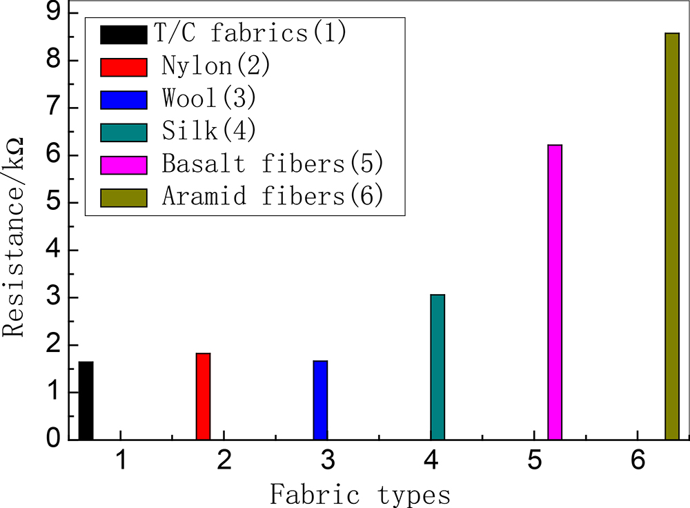
Fig. 1.
Conductivity of polypyrrole coated fabrics
Table 4.
Conductivity of polypyrrole coated fabrics after washing
| Base cloth | Blended polyester/cotton | Nylon | Wool | Silk | Basalt | Aramid |
|---|---|---|---|---|---|---|
| Resistance (kΩ) | 3.8040 | 4.2074 | 5.2350 | 8.2393 | 21.592 | 31.465 |
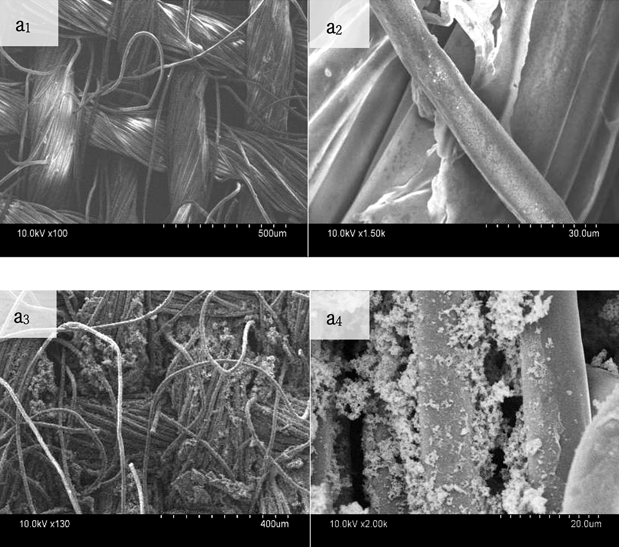
Fig. 2.
Images of electron microscopy (SEM) of polypyrrole coated terylene fabrics. Note: a1, a2 are electron microscopy images of untreated polyester-cotton blended fabrics; a3, a4 are electron microscopy images of polyester-cotton blended fabrics finished with polypyrrole
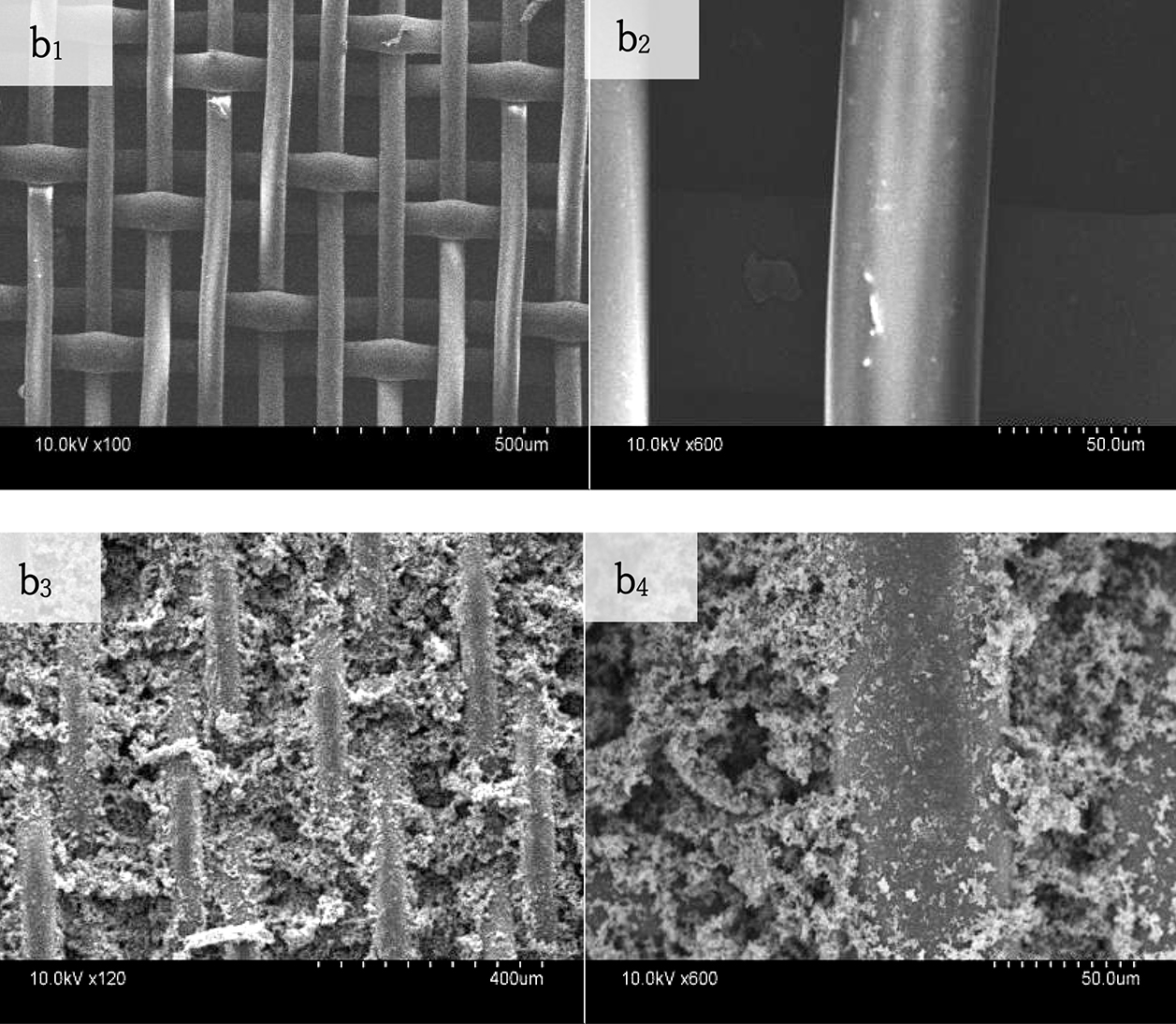
Fig. 3.
Images of electron microscopy (SEM) of polypyrrole coated nylon fabrics. Note: bt, b2 are electron microscopy images of untreated nylon fabrics; b3, b4 are electron microscopy images of nylon fabrics finished with polypyrrole
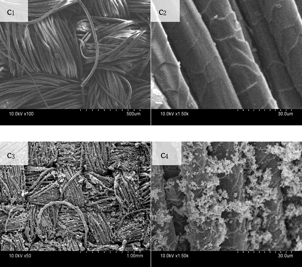
Fig. 4.
Images of electron microscopy (SEM) of polypyrrole coated wool fabrics. Note: c2, c2 are electron microscopy images of undigested wool fabrics; c3, c4 are electron microscopy images of wool fabrics finished with polypyrrole
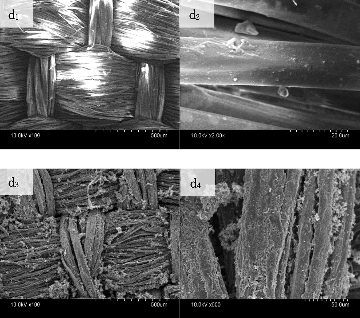
Fig. 5.
Images of electron microscopy (SEM) of polypyrrole coated silk fabrics. Note: d1, d2 are electron microscopy images of undigested silk fabrics; d3, d4 are electron microscopy images of silk fabrics finished with polypyrrole
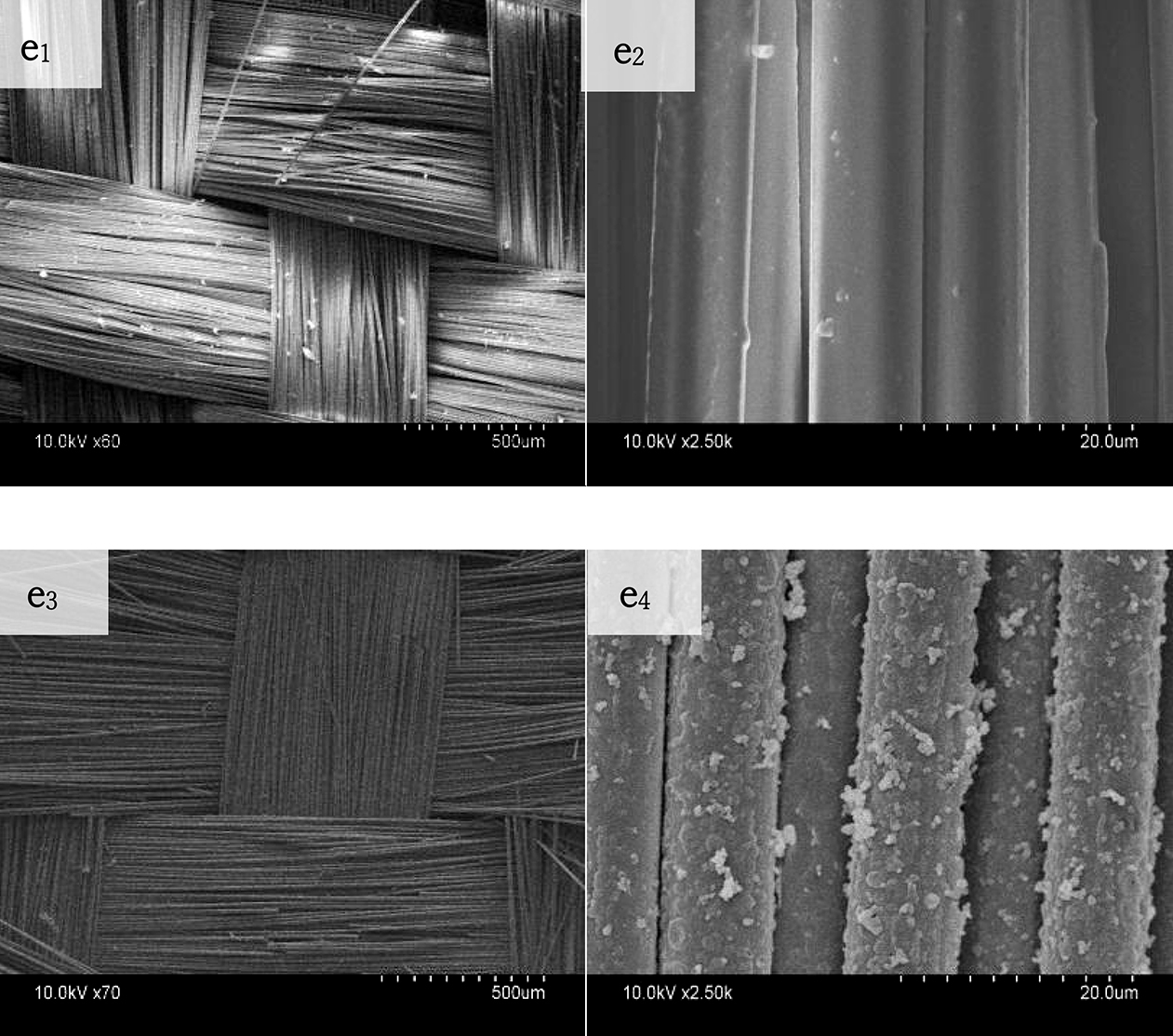
Fig. 6.
Image of electron microscopy (SEM) of polypyrrole coated basalt fabrics. Note: e1, e2 are electron microscopy images of undigested basalt fabrics; e3, e4 are electron microscopy images of basalt fabrics finished with polypyrrole
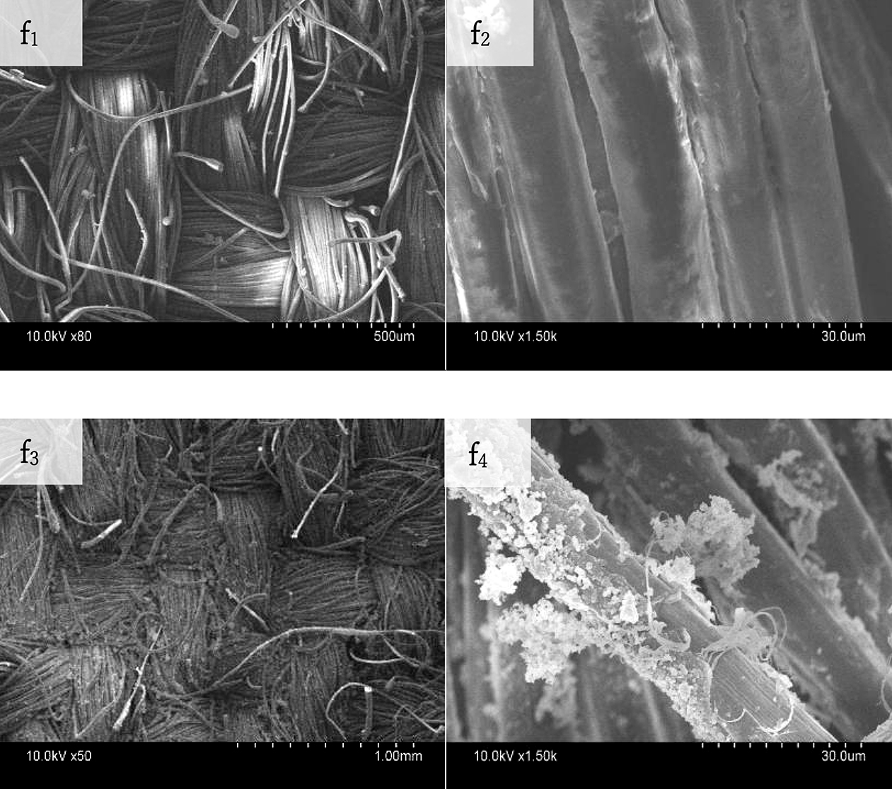
Fig. 7.
Image of electron microscopy (SEM) of polypyrrole coated aramid fabrics. Note: f1, f2 are electron microscopy images of untreated aramid fabrics; f3, f4 are electron microscopy images of aramid fabrics finished with polypyrrole