
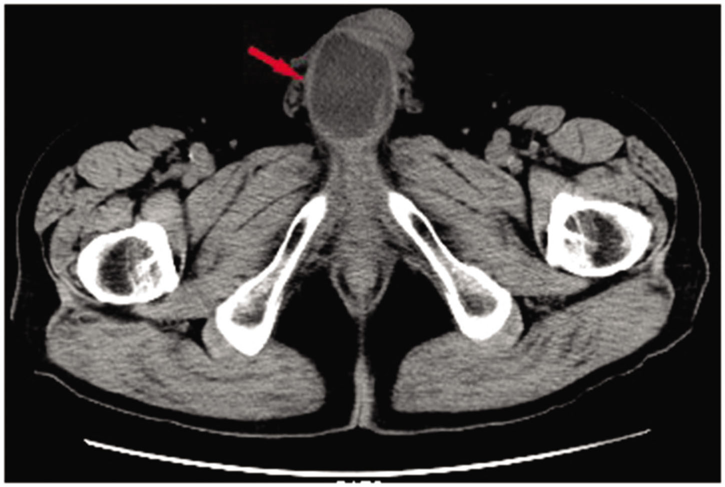
Figure 1
CT scan of a mass of 4.9 × 3.5 cm found in the cavernous body of the penis root.

Figure 2
Spiral CT 3-D image of a mass of 4.9 × 3.5 cm found in the cavernous body of the penis root.

Figure 3
T1-weighted images showing a round-like abscess in the cavernous body of the penis root.

Figure 4
T2-weighted images showing a round-like abscess in the cavernous body of the penis root.
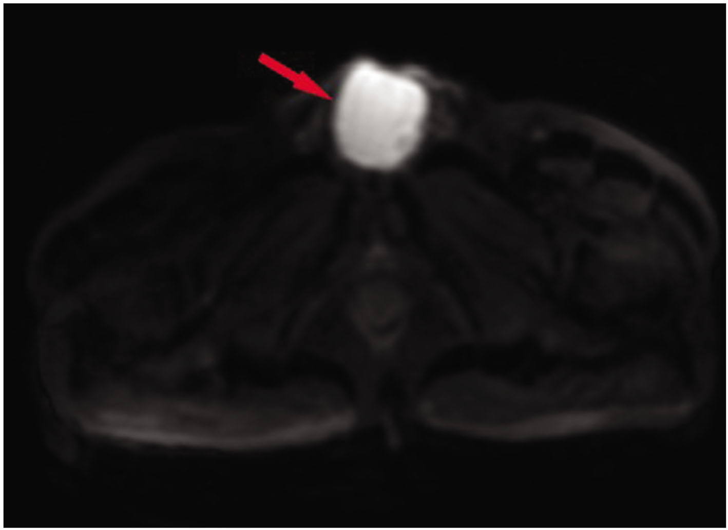
Figure 5
DWI scan showing a round-like cystic mass with high signal intensity in the cavernous body of the penis root.

Figure 6
Aspiration of about 40ml of reddish brown purulent fluid with a syringe.

Figure 7
Abscess cavity completely opened during surgery.

Figure 8
Histopathological examination showing the presence of a few inflammatory cells on the abscess wall.