
Table 1.
Schematic representation of the experiment.
| Treatments | |||||||||
|---|---|---|---|---|---|---|---|---|---|
| First stage | Control | 50 mM NaCl | 100 mM NaCl | 150 mM NaCl | 200 mM NaCl | 1 mM Gln | 2 mM Gln | 3 mM Gln | 4 mM Gln |
| Second stage | 150 mM NaCl + 1 mM Gln | 150 mM NaCl + 2 mM Gln | 150 mM NaCl + 3 mM Gln | 150 mM NaCl + 4 mM Gln | |||||
Table 2.
RAPD PCR primers and sequences.
| Primer | Primer sequence | ||
|---|---|---|---|
| RAPD2 | 5′ | ACGGTACCAG | 3′ |
| RAPD7 | 5′ | TTCCGAACCC | 3′ |
| OPPO5 | 5′ | CCCCGGTAAC | 3′ |
| OPI18 | 5′ | TGCCCAGCCT | 3′ |
| 12OPC06 | 5′ | GAACGGACTC | 3′ |
| 14OPC09 | 5′ | CTCACCGTCC | 3′ |
| 19OPC14 | 5′ | TGCGTGCTTG | 3′ |
| 21OPC16 | 5′ | CACACTCCAG | 3′ |
| OPW10 | 5′ | TCGCATCCCT | 3′ |
| 30OPAF05 | 5′ | CCCGATCAGA | 3′ |
| 25OPN01 | 5′ | CTCACGTTGG | 3′ |
| S237 | 5′ | ACCGGCTTGT | 3′ |
Table 3.
Genes, gene names, sequences and literature.
| Gene ID | Gene name | Primer sequences (5→3’) | Reference |
|---|---|---|---|
| SOD | Superoxide dismutase | F TTCCTCCAGCATTCCCAGTG | Ghodke et al. (2020) |
| R ATGGCTTGACACATGGTGCT | |||
| SOD-2 | Superoxide dismutase 2, mitochondrial | F GGCGAAGCAAACAGCCTCAT | Ji et al. (2021) |
| R AGTATCGCCGAACGAGTGGA | |||
| AO | L-ascorbate oxidase-like | F TGATGTTTGTGCTGTCTTTCGG | Ghodke et al. (2020) |
| R ACCGTGAAAGTGTTTGTGCT | |||
| POLD1 | DNA Polymerase Delta 1, catalytic subunit | F AGACGACTCGCTGTGTATTGCT | Lyu et al. (2020) |
| R CCAGTAACTCGTGCCATCTCCA | |||
| Chape | Chaperone | F TCCTGGCAAGTCTGCTTTGA | Ghodke et al. (2020) |
| R GGCTCTAATTCCTCGCGTTT | |||
| HSP21 | 21 kDa protein | F TAGATGGATGGCGAACTCGG | Ghodke et al. (2020) |
| R TCTTCTTCTTCGCACTCCT | |||
| β-Actin | β-actin | F TCCTAACCGAGCGAGGCTACAT | Sun et al. (2013) |
| R GGAAAAGCACTTCTGGGCACC | |||
| 18S | 18S ribosomal RNA | F GAATGACTCCTGGCAATG | Liu et al. (2015) |
| R GATTGGAATGACGCTATACA |
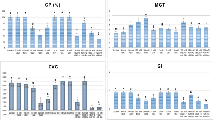
Figure 1.
Changes in GP, MGT, CVG, and GI caused by salt stress and Gln treatments. Statistically significant differences between treatments are shown in the bar graphs with different letters. According to one-way ANOVA (Tukey test), statistically significant differences were determined between the experimental groups at the p ≤ 0.05 level. ANOVA, analysis of variance; CVG, coefficient of velocity of germination; GI, germination index; GP, germination percentage; MGT, mean germination time.
Table 4.
Changes in the vegetative growth of onions at different Gln concentrations under salt-stress and non-stress conditions.
| Treatments | SL (mm) | RL (mm) | SFW (g) | RFW (g) | SDW (g) | RDW (g) | TLN (no.) |
|---|---|---|---|---|---|---|---|
| Control | 300.78 ab | 335.30 a | 4.7717 bc | 5.3486 a | 0.2974 b | 0.2578 b | 5.4 c |
| 50 mM NaCl | 200.50 abc | 174.67 b | 3.3849 c | 1.8111 bcd | 0.2423 b | 0.1269 cd | 6.0 c |
| 100 mM NaCl | 176.14 bcd | 99.98 bcd | 3.0034 c | 0.9761 cd | 0.3421 b | 0.0514 de | 4.7 c |
| 150 mM NaCl | 55.37 de | 54.16 cd | 1.6138 c | 0.7861 cd | 0.1272 b | 0.0980 de | 4.3 c |
| 200 mM NaCl | 35.96 e | 30.95 d | 0.6784 c | 0.2976 d | 0.1009 b | 0.0250 e | 3.6 c |
| 1 mM Gln | 265.67 ab | 78.67 bcd | 5.6750 abc | 2.0207 bcd | 0.3935 b | 0.1229 cd | 6.7 bc |
| 2 mM Gln | 316.00 a | 131.33 bc | 11.1293 a | 3.1972 b | 5.0274 a | 0.2022 bc | 11.6 ab |
| 3 mM Gln | 269.00 ab | 70.00 cd | 10.4772 ab | 2.8864 bc | 3.4157 a | 0.9267 a | 13.7 a |
| 4 mM Gln | 243.67 ab | 68.67 cd | 6.4551 abc | 2.2977 bcd | 4.5882 a | 0.2567 b | 6.6 bc |
| 150 mM NaCl+ 1 mM Gln | 64.67 de | 79.67 bcd | 1.3471 c | 1.8282 bcd | 0.1246 b | 0.1017 de | 3.7 c |
| 150 mM NaCl+ 2 mM Gln | 65.33 de | 78.00 cd | 1.6087 c | 1.3310 bcd | 0.1228 b | 0.1293 cd | 6.0 c |
| 150 mM NaCl+ 3 mM Gln | 81.67 cde | 84.67 bcd | 1.7282 c | 1.8951 bcd | 0.1594 b | 0.1341 cd | 6.6 bc |
| 150 mM NaCl+ 4 mM Gln | 77.67 cde | 64.00 cd | 0.8713 c | 1.1304 bcd | 0.1030 b | 0.1166 cde | 5.7 c |
| ** | ** | ** | ** | ** | ** | ** |
1 Statistically, the differences between the applications are shown with different letters (Tukey). ** p ≤ 0.001, * p ≤ 0.005, ns: not significant. Gln: glutamine, SL: shoot length, RL: root length, SFW: shoot fresh weight, RFW: root fresh weight, SDW: shoot dry weight; RDW: root dry weight; TLN: total leaf number.
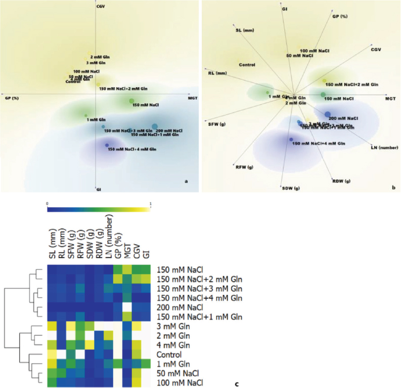
Figure 2.
Linear projection of the distribution of applications according to plant growth and germination values and visualisation of the classification of applications according to the change in plant characteristics with heat map. (A) Linear projection according to germination attributes, (B) linear projection according to germination and vegetative attributes (C) heat map. By using the principal component analysis data in linear projection (A and B), a two-dimensional projection is presented in which different applications are best separated according to variables. As it can be followed from the scale on the heat map (C), the change of colours gives information about the effect of the applications on the variables. The lowest values are shown in dark blue. The change of colour towards white in the heat map showed that the values increased.
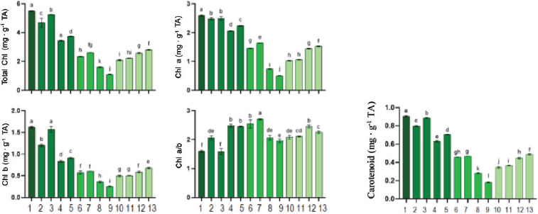
Figure 3.
Total Chl, Chl a, Chl b, Chl a/b and carotenoid amounts determined in leaf tissues of control and experimental groups of onion. Data are given as mean ± standard deviation, n = 5. The averages shown with different letters on the graph are statistically different from each other. One-way ANOVA, TUKEY HSD test, p ≤ 0.05 (1: Control, 2: 1 mM Gln, 3: 2 mM Gln, 4: 3 mM Gln, 5: 4 mM Gln, 6: 50 mM NaCl, 7: 100 mM NaCl, 8: 150 mM NaCl, 9: 200 mM NaCl, 10: 150 mM NaCl + 1 mM Gln,11: 150 mM NaCl + 2 mM Gln, 12: 150 mM NaCl + 3 mM Gln, 13: 150 mM NaCl + 4 mM Gln). ANOVA, analysis of variance; Chl a, chlorophyll a; Chl a/b, chlorophyll a/b ratio; Chl b, chlorophyll b; total Chl, total chlorophyll.
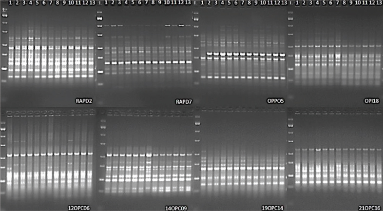
Figure 4.
Gel image of the primers included in the analysis. (1: Control, 2: 1 mM Gln, 3: 2 mM Gln, 4: 3 mM Gln, 5: 4 mM Gln, 6: 50 mM NaCl, 7: 100 mM NaCl, 8: 150 mM NaCl, 9: 200 mM NaCl, 10: 150 mM NaCl + 1 mM Gln, 11: 150 mM NaCl + 2 mM Gln, 12: 150 mM NaCl + 3 mM Gln, 13: 150 mM NaCl + 4 mM Gln). L (Ladder): Fermantas zip ruler DNA ladder 100 bp.
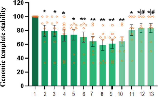
Figure 5.
GTS in the control and treatment groups. n = 8, data mean ± standard error. Comparison of treatment groups compared to control, data are statistically different, one-way ANOVA test, *p ≤ 0.05, **p ≤ 0.01; #comparative applications vs. 150 mM NaCl, data are statistically different, independent groups t-test, p ≤ 0.05 (1: Control, 2: 1 mM Gln, 3: 2 mM Gln, 4: 3 mM Gln, 5: 4 mM Gln, 6: 50 mM NaCl, 7: 100 mM NaCl, 8: 150 mM NaCl, 9: 200 mM NaCl, 10: 150 mM NaCl + 1 mM Gln,11: 150 mM NaCl + 2 mM Gln,12: 150 mM NaCl + 3mM Gln, 13: 150 mM NaCl + 4mM Gln). ANOVA, analysis of variance; GTS, genomic template stability.
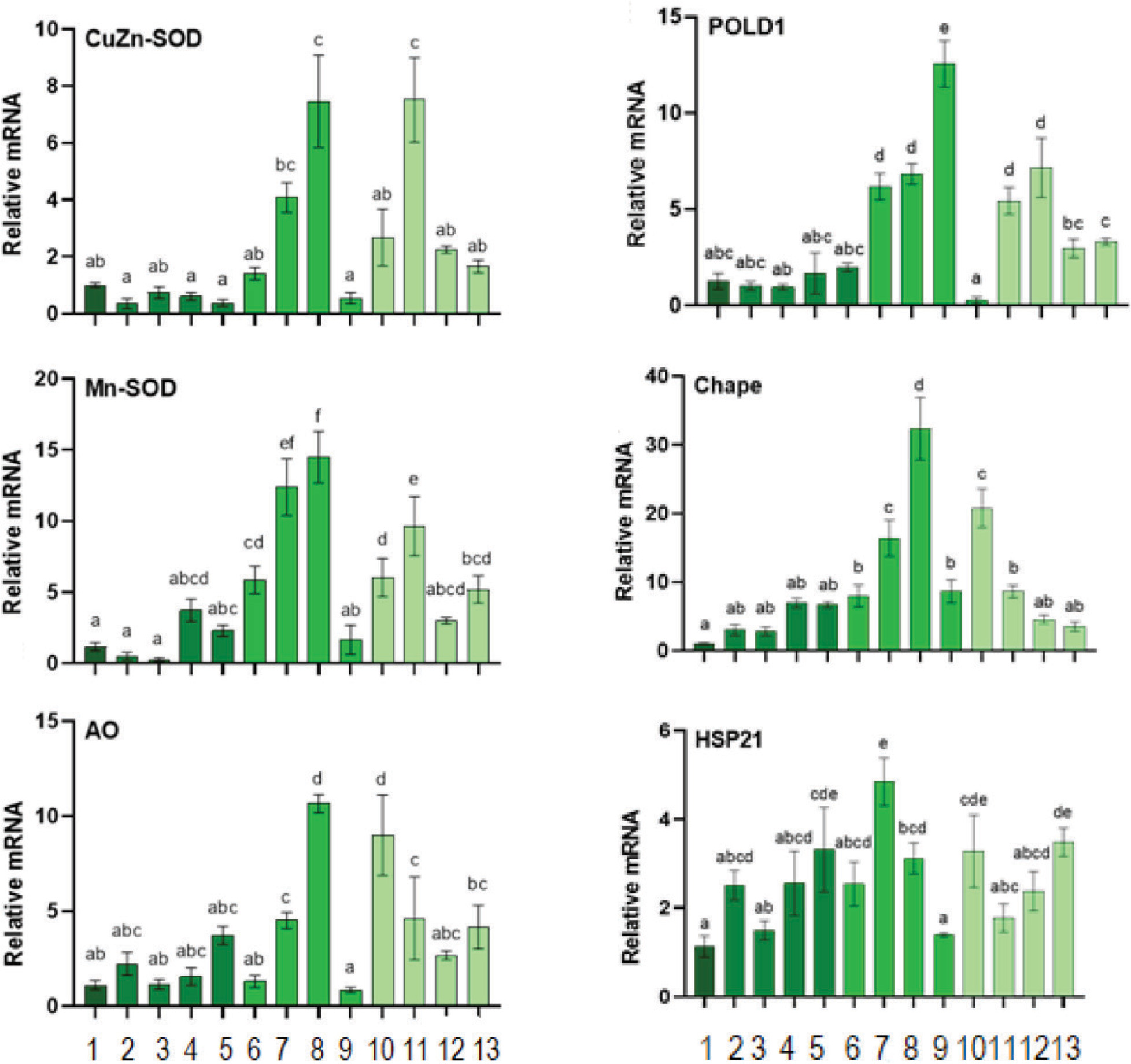
Figure 6.
Relative fold increased values of antioxidant defence genes CuZn-SOD, Mn-SOD, AO, DNA damage repair gene POLD1, heat-shock molecular chaperone CHAPE and HSP21 gene expressions in leaf tissues in experimental groups (data are β-actin and normalised to 18S mRNA level by the multiple control method). Data are given as mean ± standard error, n = 5. The averages shown with different letters on the graph are statistically different from each other. One-way ANOVA, Tukey HSD test, p ≤ 0.05 (1: Control, 2: 1 mM Gln, 3: 2 mM Gln, 4: 3 mM Gln, 5: 4 mM Gln, 6: 50 mM NaCl, 7: 100 mM NaCl, 8: 150 mM NaCl, 9: 200 mM NaCl, 10: 150 mM NaCl + 1 mM Gln,11: 150 mM NaCl + 2 mM Gln,12: 150 mM NaCl + 3mM Gln, 13: 150 mM NaCl + 4 mM Gln). ANOVA, analysis of variance.