
Table 1
Primers used for detection of CVB by RT-PCR and colourimetric RT-LAMP techniques in this study.
| Type of detection | Primer name | Sequence (5′-3′) | Target gene(s) | Ta*** (°C) | Reference |
|---|---|---|---|---|---|
| PCR | CVB-F1 CVB-R1 | AGTCACAATGCCTCCCAAAC CATACCTTTCTTAGAGTGCTATGCT | TGB3** + CP | 54 | Guan et al. (2017) |
| PCR and sequencing | CVB-up CVB-dw | TAGGTTGTGGAGTGGTTACA ATCTTCACAATGACATCCAT | CP | 56 | Lin et al. (2005) |
| LAMP | FIP* BIP F3 B3 | CCTGCTCACGCTCTCGTTCCCAGCTCGAACAGCGGAAG AGATGAACTCCAATGCCCCAGCTAGTGCCGCGAGTTGTGT ACCAACTCCACCTCCACC TCGTCTTGCTCTCTCCTCAG | TGB3 + CP | 63–68 | This study |
BIP, backward inner primer containing B1c + B2; B3, backward primer; CP, coat protein gene; CVB, chrysanthemum virus B; F3, forward primer; LAMP, loop-mediated isothermal amplification; PCR, polymerase chain reaction; RT-PCR, reverse transcription polymerase chain reaction.
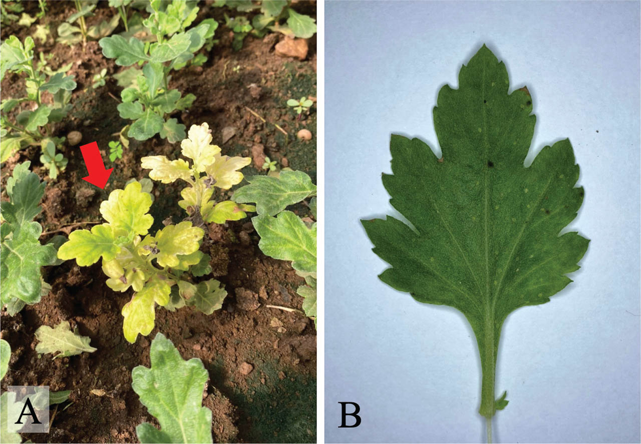
Figure 1
Symptoms observed on chrysanthemum leaves collected from Chiang Mai Province of Thailand that were positive to CVB detection by using RT-PCR. (A) Chlorosis and yellowing symptoms (the red arrow). (B) The mild mottling symptom. CVB, chrysanthemum virus B; RT-PCR; reverse transcription polymerase chain reaction.
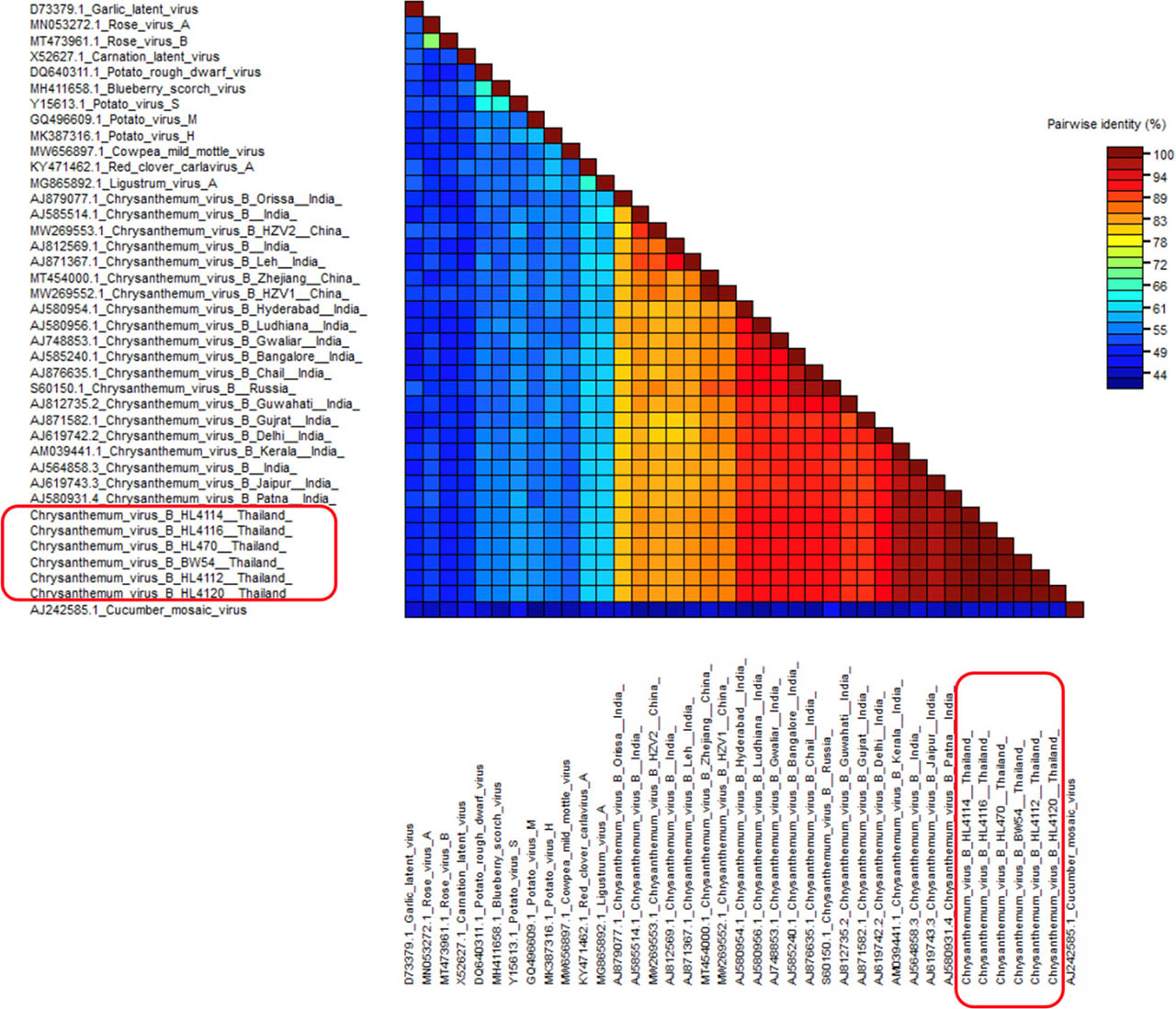
Figure 2
Colour-coded pairwise identity matrix generated from 35 virus sequences consisting of CVB isolates, other members of the genus Carlavirus and CMV of the genus Cucumovirus that was used as the outgroup. CVB isolates in this study were indicated by red boxes. The pairwise identity matrix was performed in the SDT version 1.2 program. The colour of the cell indicates the percentage identity score between two sequences (left and bottom axis) according to the colour-coded cells mean to each cell in the SDT matrix of the Figure 2. CMV, cucumber mosaic virus; CVB, chrysanthemum virus B; SDT, sequence demarcation tool.
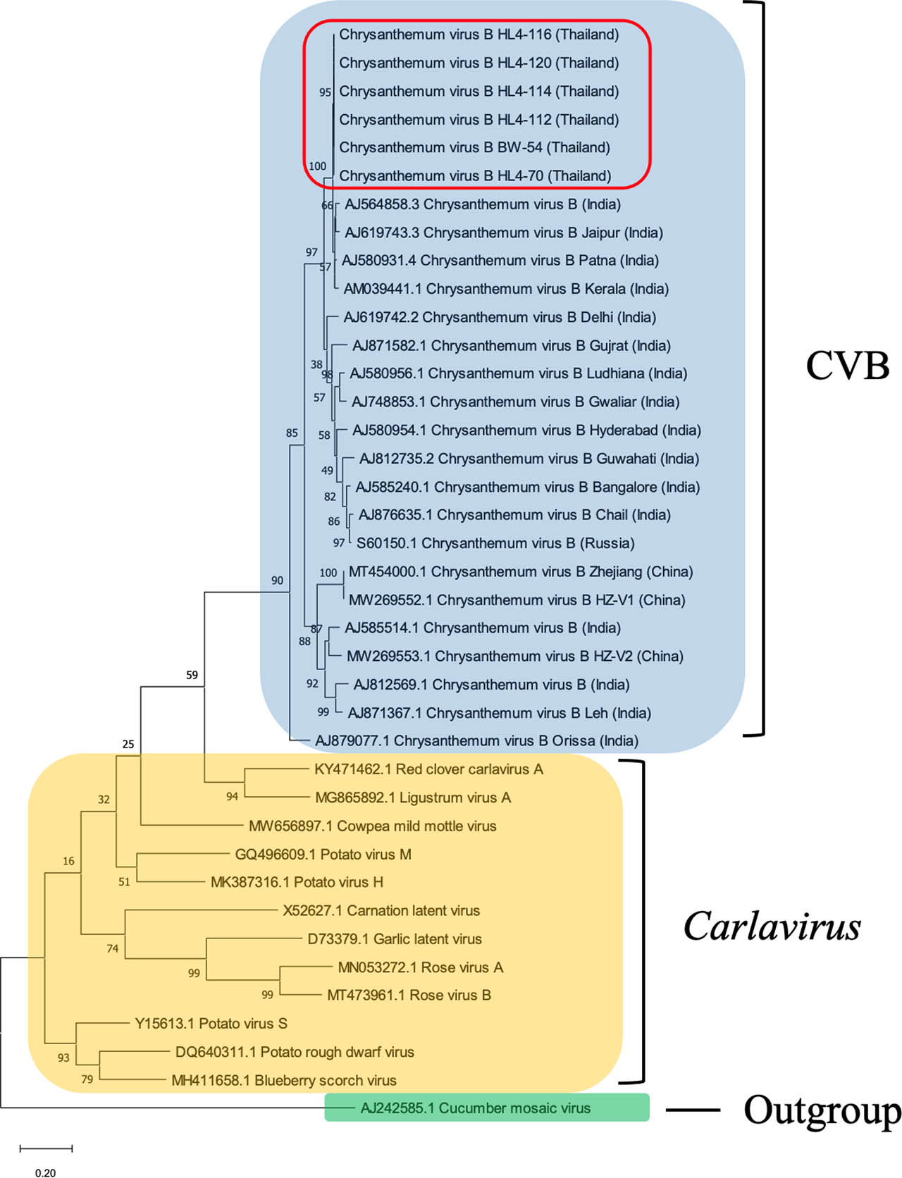
Figure 3
The phylogenetic relationship of CVB-BW-54 and CVB-HL4-70 (the red box) and members of the genus Carlavirus based on nucleotide sequences of the CP gene. CMV (accession №. AJ242585.1) was used as the outgroup. Multiple sequence alignments and construction of the phylogenetic tree were generated by ClustalW by the ML method with 1,000 replicates of bootstrap values performed by MEGA X program. CMV, cucumber mosaic virus; CP, coat protein; ML, maximum likelihood.
Table 2
Symptoms induced by CVB on indicator plants after mechanical inoculation.
| Indicator plants | Symptoms | |
|---|---|---|
| Inoculated leaf | Upper leaf | |
| Chenopodium amaranticolor | CS | – |
| C. quinoa | CS | – |
| Chrysanthemum × morifolium | – | Y |
| Nicotiana benthamiana | – | M, Ma |
| N. glutinosa | NS | – |
| N. tabacum cv. Xanthi | – | M, Ma |
| N. tabacum cv. Samsun | NS | – |
| Petunia × hybrida | – | M, Ma |
| Vigna uniculata | – | – |
[i] CS: chlorotic spot; CVB, chrysanthemum virus B; M, mosaic; Ma, malformation; NS, necrotic spot; Y, yellowing; –, no symptom.
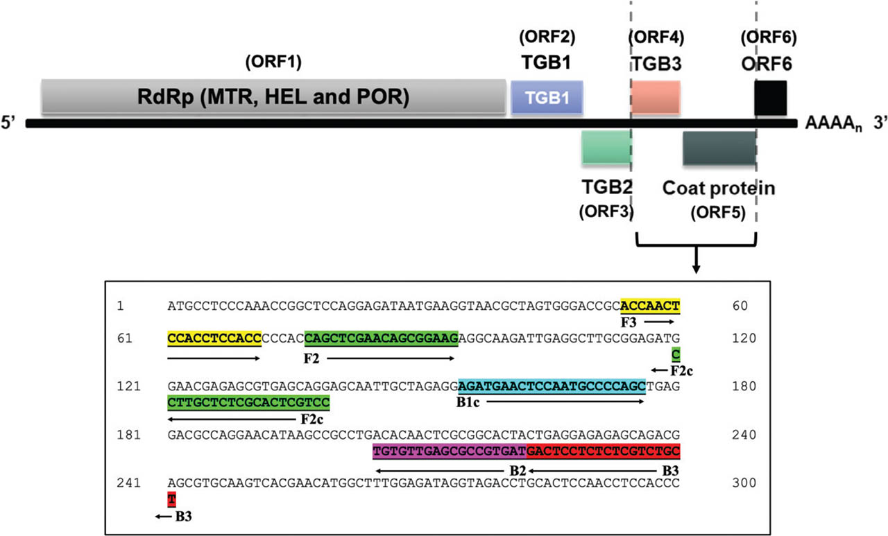
Figure 4
Diagram of LAMP primers’ attachment on the partial ORF4 (TGB3 gene) and ORF5 (CP gene) of CVB genome. All four LAMP primers including FIP (containing F2 and F2c), BIP (containing B2 and B1c), F3 and B3 were highlighted with different colours. BIP, backward inner primer; B3, backward primer; CVB, chrysanthemum virus B; FIP, forward inner primer; F3, forward primer; LAMP, loop-mediated isothermal amplification.
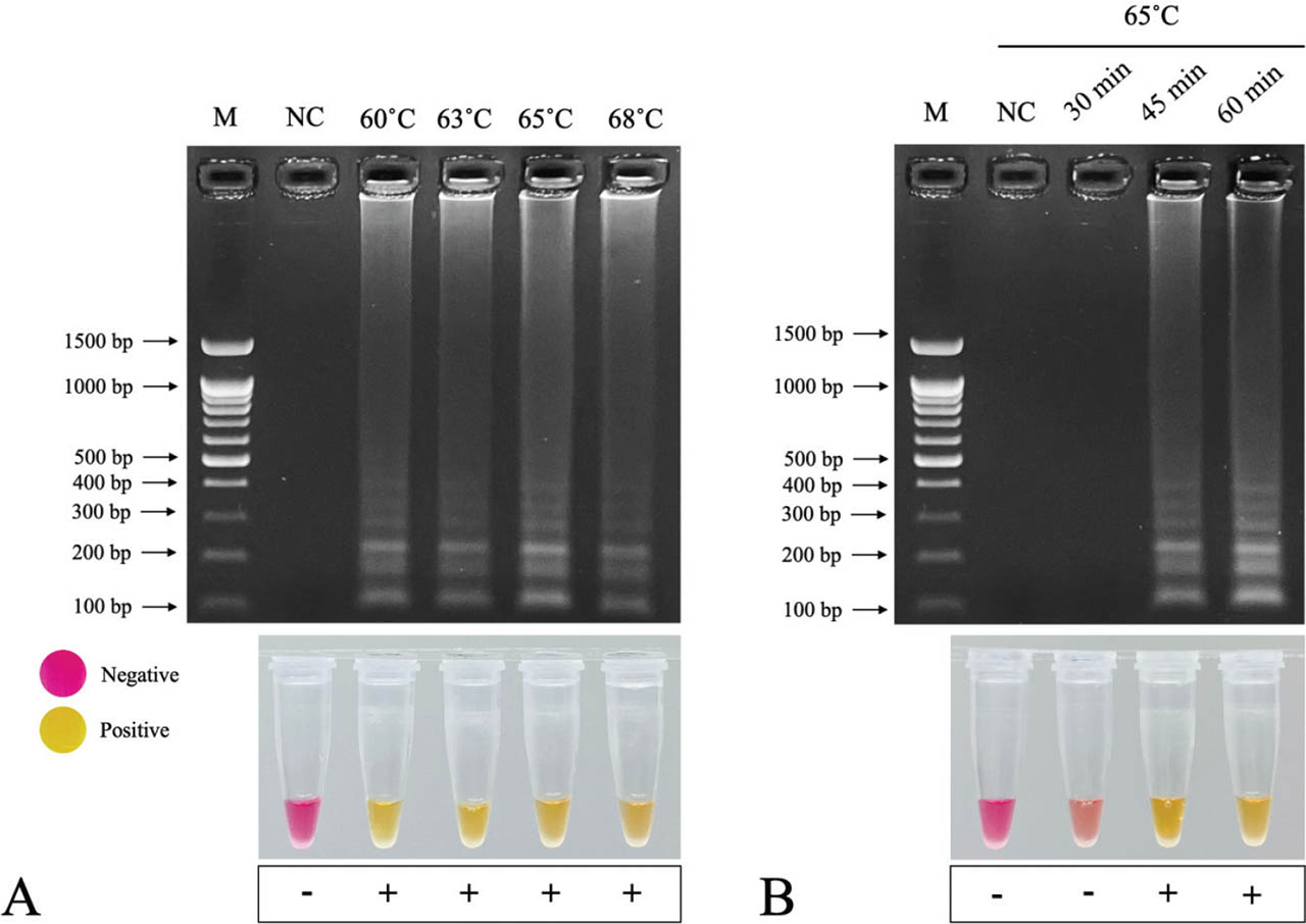
Figure 5
Optimisation of RT-LAMP for detection of CVB. (A) Optimisation of temperature at 60 °C, 63 °C, 65 °C and 68 °C for 60 min. (B) Optimising time of incubation at 65 °C for 30 min, 45 min and 60 min. CVB, chrysanthemum virus B; M, DNA ladder; NC, negative control (nuclease-free water).
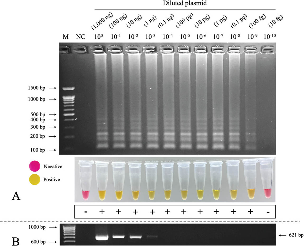
Figure 6
Sensitivity test of LAMP in the detection of CVB compared with RT-PCR detection. (A) LAMP-AGE (top) with the LOD at 10−9 diluted plasmid and colourimetric LAMP (bottom). (B) PCR with LOD at 10−3 diluted plasmid. AGE, agarose gel electrophoresis; CVB, chrysanthemum virus B; LAMP, loop-mediated isothermal amplification; LOD, limits of detection; M, DNA ladder; NC, negative control (nuclease-free water); PCR, polymerase chain reaction; RT-PCR, reverse transcription polymerase chain reaction.
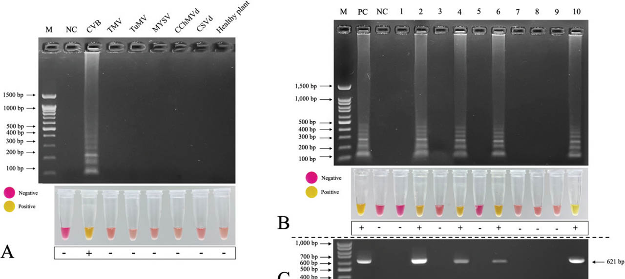
Figure 7
Specificity assay and evaluation of colourimetric RT-LAMP for detection of CVB. (A) The LAMP product was observed only in the lane of CVB. (B) Evaluation of colourimetric RT-LAMP for detection of CVB from random chrysanthemum samples. (C) RT-PCR detection. CChMVd, chrysanthemum chlorotic mottle viroid; CSVd, chrysanthemum stunt viroid; CVB, chrysanthemum virus B; LAMP, loop-mediated isothermal amplification; Lanes 1–10, chrysanthemum samples; M, DNA ladder; MYSV, melon yellow spot virus; NC, negative control (nuclease-free water); PC, positive control; RT-PCR, reverse transcription polymerase chain reaction; TMV, tobacco mosaic virus; TuMV, turnip mosaic virus.