
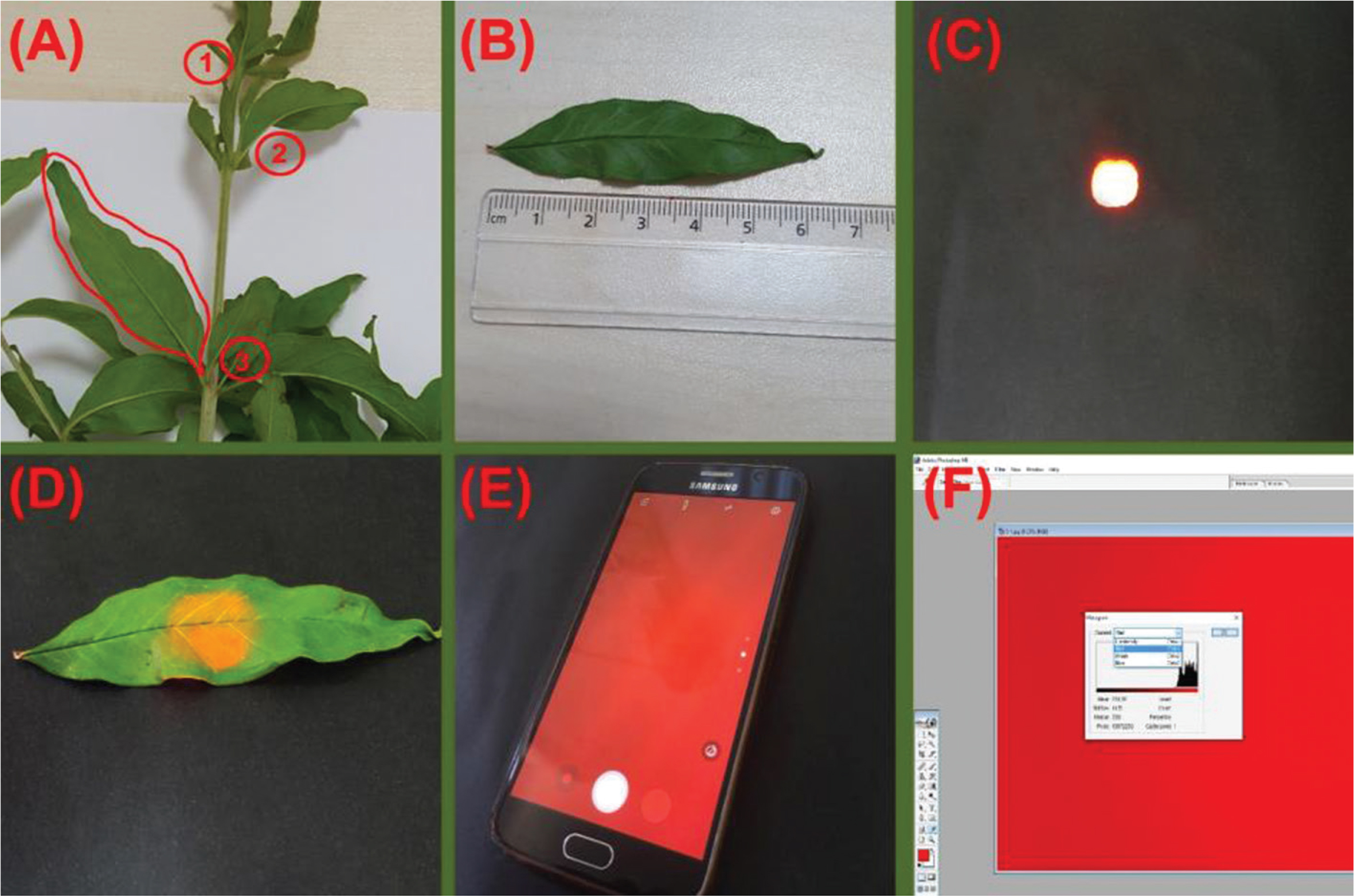
Figure 1
Experimental methodology and image acquisition: (A) selection of the leaf samples, (B): determination of the mid-point of the leaves, (C) a view of the red LED light from the closed box, (D) insertion of the leaf onto the closed box’s hole, (E) capturing of the contact image with a smartphone and (F) reading the RGB values of the images by using the histogram function of the Photoshop ME 7.0.
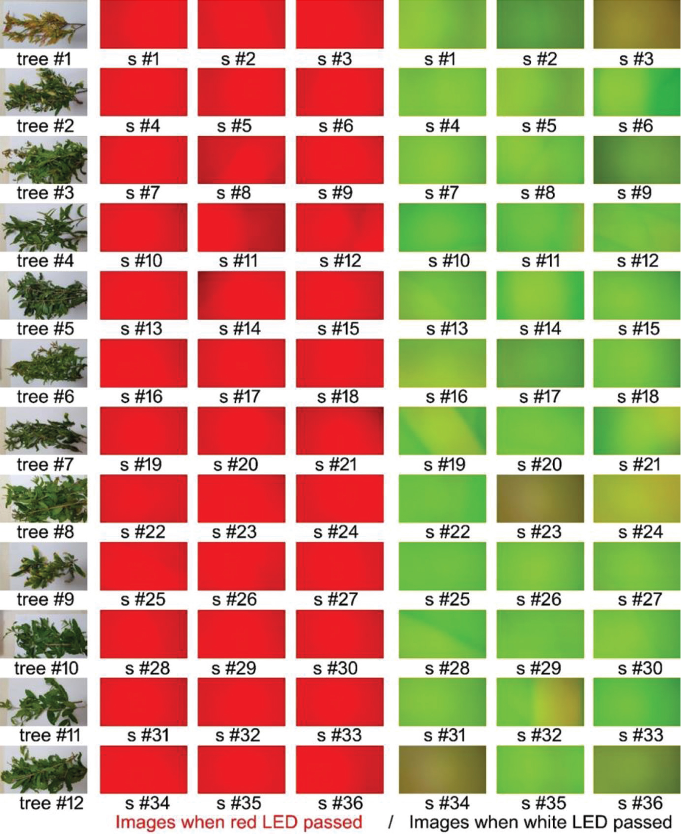
Figure 2
Captured leaf images with a smartphone when red LED passed (three red images from the left side) and when white LED passed (three green images from the right side).
Table 1
Correlation between the RGB colours and combination indices and leaf chlorophyll contents (for both mg ∙ g−1 and mg ∙ MW−1) of a pomegranate tree
| RGB colours and combination indices | Red LED passed | White LED passed | ||
|---|---|---|---|---|
| Chl (mg ∙ g−1) | Chl (mg ∙ MW−1) | Chl (mg ∙ g−1) | Chl (mg ∙ MW−1) | |
| R value | −0.282 | −0.552** | −0.225 | −0.257 |
| G value | 0.113 | 0.914** | 0.210 | 0.460** |
| B value | 0.146 | 0.893** | −0.382* | −0.364* |
| NRI | −0.154 | −0.914** | −0.195 | −0.424** |
| NGI | 0.148 | 0.918** | 0.344* | 0.476** |
| NBI | 0.172 | 0.897** | −0.384* | −0.371* |
| GMR | 0.280 | 0.571** | 0.279 | 0.484** |
| GMB | 0.000 | 0.756** | 0.287 | 0.456** |
| GRRI | 0.146 | 0.917** | 0.233 | 0.450** |
| GRVI | 0.144 | 0.916** | 0.302 | 0.475** |
| DGCI | 0.280 | 0.574** | −0.014 | 0.009 |
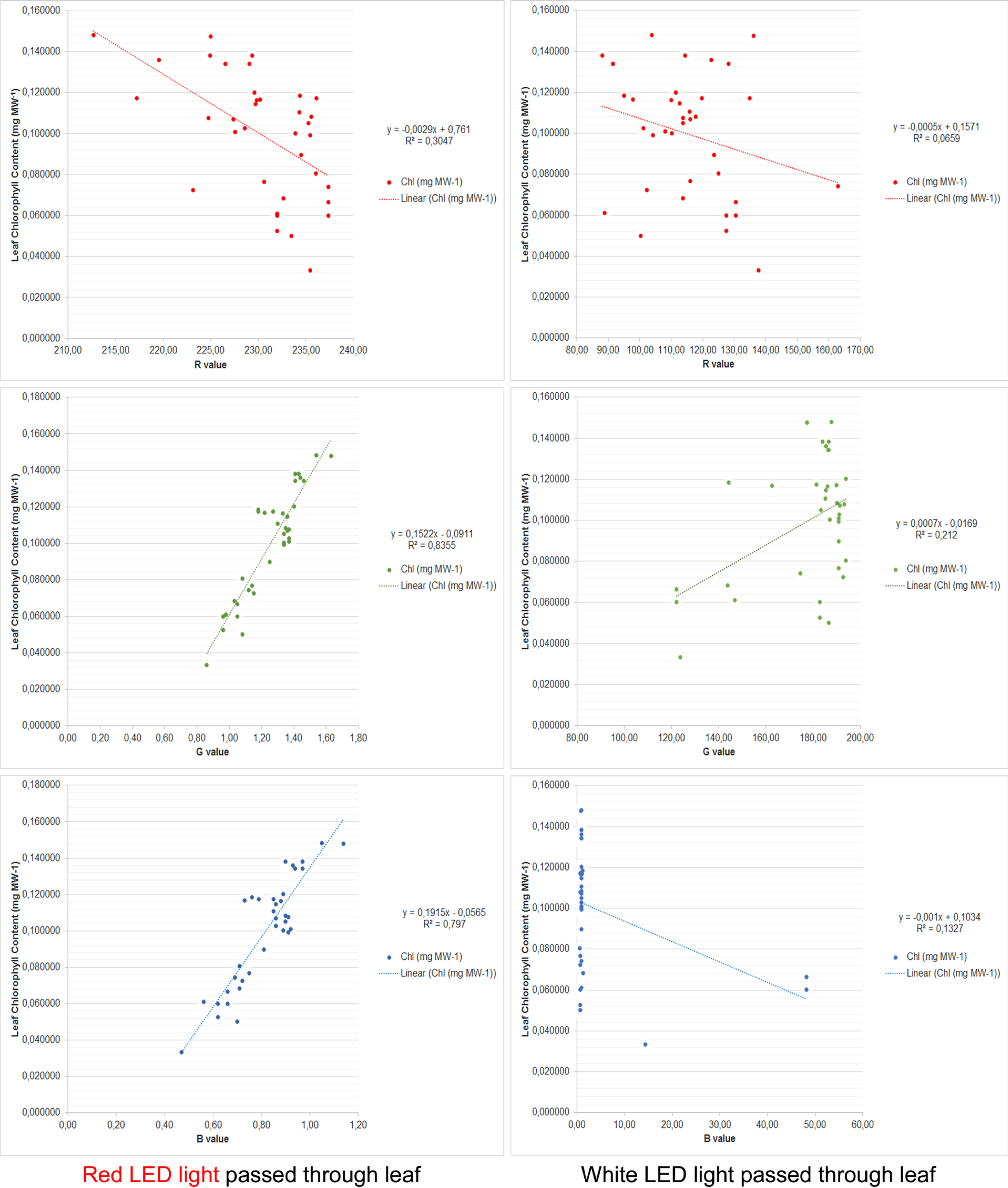
Figure 3
Stepwise linear regression analysis for leaf Chl content measured analytically in laboratory and the RGB values based on contact imaging with smartphone.