
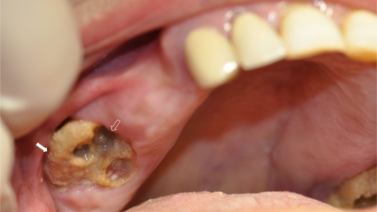
Figure 1:
Osteonecrosis of the maxilla, presenting as exposed necrotic bone (compact arrow), after a dental extraction. Sockets without teeth (phantom sockets) (transparent arrow), are seen on the necrotic bone. Patient was asymptomatic at presentation.
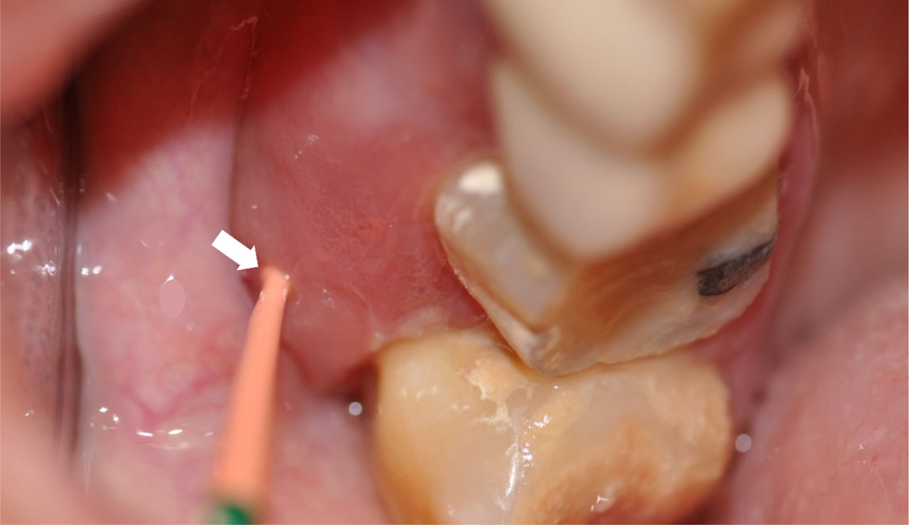
Figure 2:
Osteonecrosis on the maxilla was diagnosed after probing necrotic bone through a fistula (arrow). A plastic material (gutaperka) was inserted into the fistula to better disclose the fistula. Patient presented with pain and swelling.
Table 1:
MRONJ Stages, American Association of Oral and Maxillofacial Surgeons 2014
| Stage 0 | Stage 1 | Stage 2 | Stage 3 |
|---|---|---|---|
| Clinical definitions | |||
|
|
|
|
| Radiographic features | |||
|
|
|
|
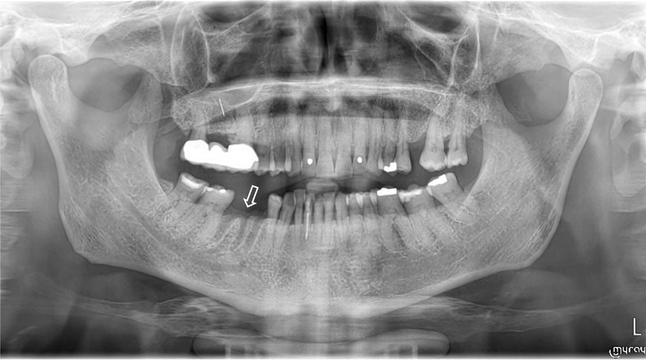
Figure 3:
Panoramic radiograph of a cancer patient, who received antiresorptives and presented with a non-healing socket (phantom socket) two months after the dental extraction of the mandibular molar tooth. Patient was diagnosed with MRONJ stage 2.
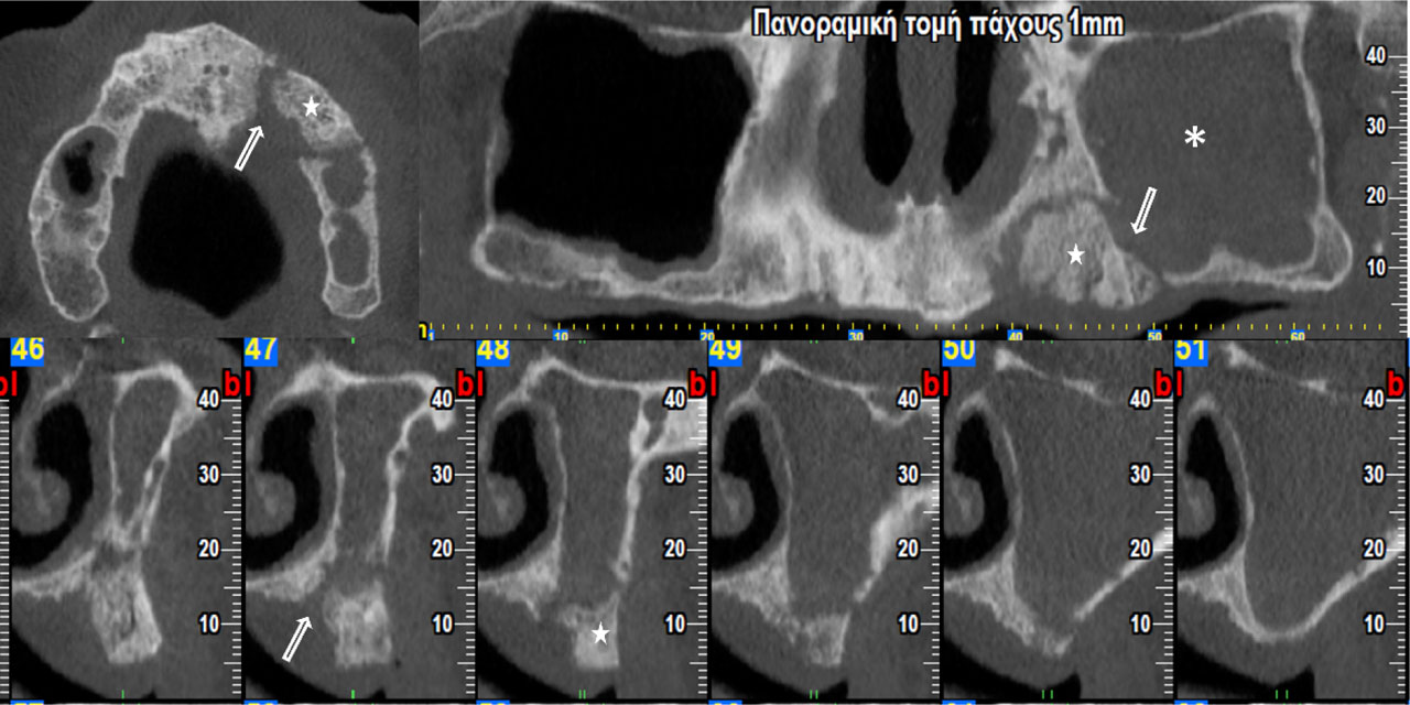
Figure 4:
Cone Beam Computed Tomography of a cancer patient, who received antiresorptives. Osteolytic areas (arrows) and bone sequestrum (stars) are observed in the maxilla. Fullness of left sinus is also seen (asterisks). Patient was diagnosed with MRONJ stage 3.