
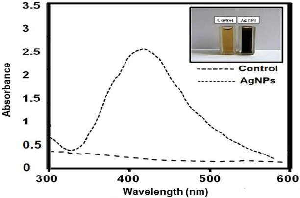
Figure 1
Spectrophotometric analysis of biosynthesized silver nanoparticles. Control - Initial mixture containing Lactobacillus supernatant and 1mM silver nitrate in 1:1 ratio. AgNPs - Color change observed of reaction mixture containing Lactobacillus supernatant and 1mM silver nitrate in 1:1 ratio
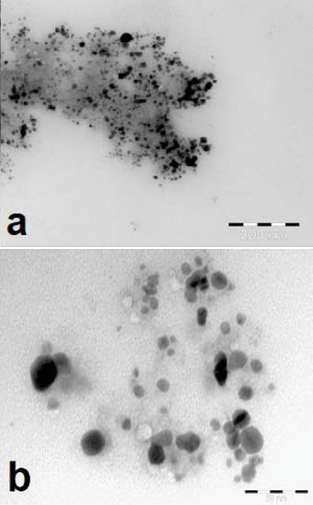
Figure 2
Transmission Electron Microscopy (TEM) micrographs of silver nanoparticles synthesized with culture filtrate of Lactobacillus sp. strain LCM5 at different magnifications (a) 30000x, (b) 200000x.
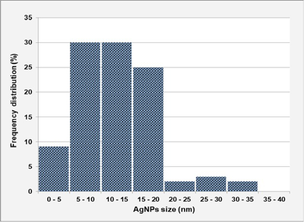
Figure 3
The particles size distribution of synthesized AgNPs.
Table 1
Antimicrobial activity of silver nanoparticles
| Test microorganism | Inhibition zone (mm) Ag NO3 | Inhibition zone (mm) Ag NPs |
|---|---|---|
| Aspergillus flavus | 9.00 ± 1.06c* | 12.39 ± 0.61c |
| Aspergillus ochraceus | 7.00 ± 0.37d | 12.86 ± 0.78c |
| Penicillium expansum | 11.00 ± 0.46b | 15.87 ± 1.01b |
| Chromobacterium violaceum | 16.00 ± 0.67a | 18.00 ± 0.69a |
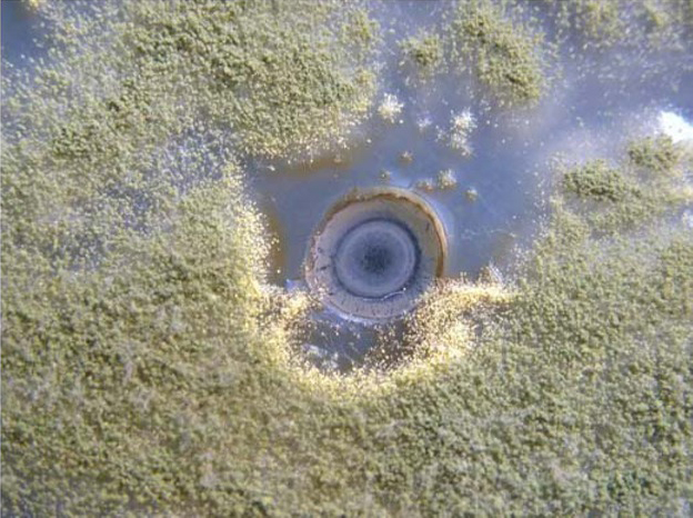
Figure 4
Antifungal effect of AgNPs against Aspergillus flavus.
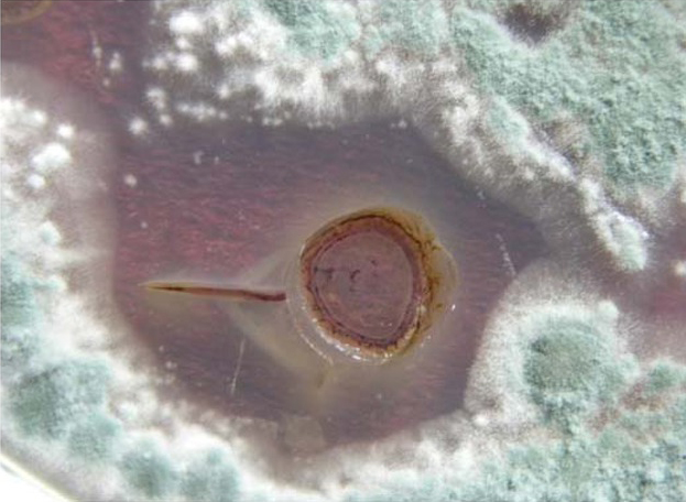
Figure 6
Antifungal effect of AgNPs against Penicillium expansum.
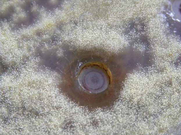
Figure 5
Antifungal effect of AgNPs against Aspergillus ochraceus.
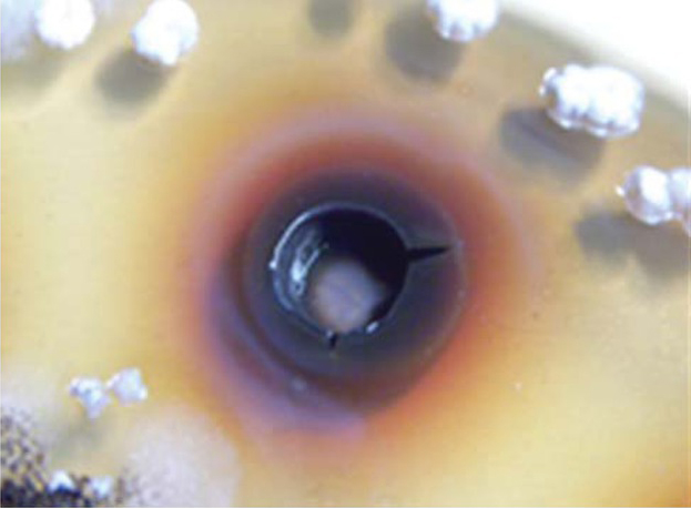
Figure 7
Antibacterial effect of AgNPs against Chromobacterium violaceum.