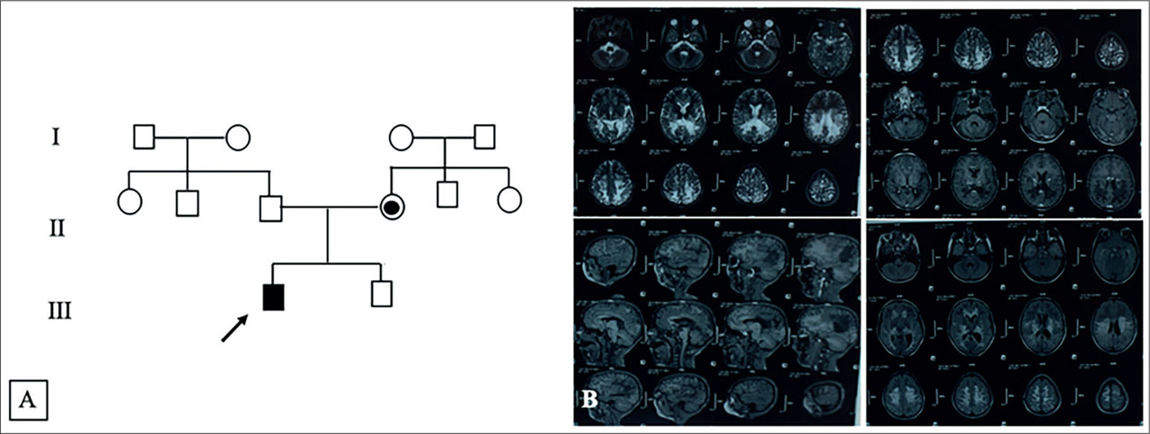
Figure 1.
Family pedigree (A) with the Brain MRI Images of the patient showing signal abnormalities in the parieto-temporo-occipital white matter (B).
Table 1.
Table summarizing the clinical signs of the patient.
| Patient symptoms | |
|---|---|
| Gender | Male |
| Age at the diagnosis | 12 |
| Age at onset | 10 |
| Initial symptoms | Academic difficulties |
| Spinal symptoms | Spastic paraparesis |
| Peripheral neuropathy | Present |
| Cognitive impairment | Present |
| Sphincter dysfunction | Present |
| hypoadrenocorticism | Present |
| Muscle strengh | Increased |
| Tendon reflexes | Exaggerated |
| Hoffman sign | Positive |
| Babinski sign | Positive |
| Sensory | Normal |
| Cerebral involvement in MRI | Present |
| Spinal involvement in MRI | Absent |
| Disease progression | Rapid |
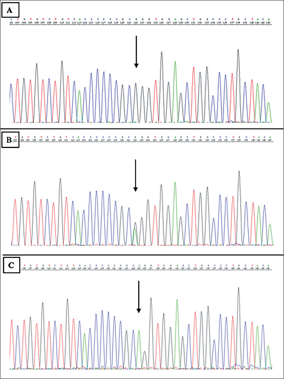
Figure 2.
Electropherograms showing the pathogenic variant in the patient in a hemizygous state (A), in a heterozygous state in the mother (B), and the absence of the pathogenic variant in the healthy brother (C).