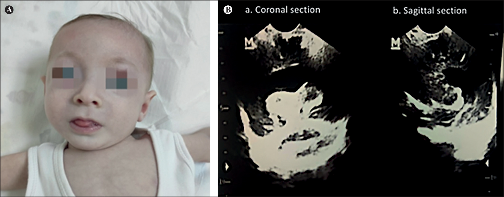
Figure 1.
(A) Seven months old infant presenting with microcephaly and mild facial dysmorphic features, and (B) coronal (a) and sagittal (b) section of trans-fontanelle ultrasound examination revealing a single ventricle.

Figure 2.
1,5 T MRI showing mega cisterna magna, rudimentary anterior falx cerebri without formed fronto-parietal interhemispheric fissure, absence of septum pellucidum with agenesis of the corpus callosum, thinning of cerebrum, dysplastic unfused thalamus, rudimentary third brain ventricle, rudimentary temporal and occipital horns and cortical polymorphia with macrogyria and microgyria.
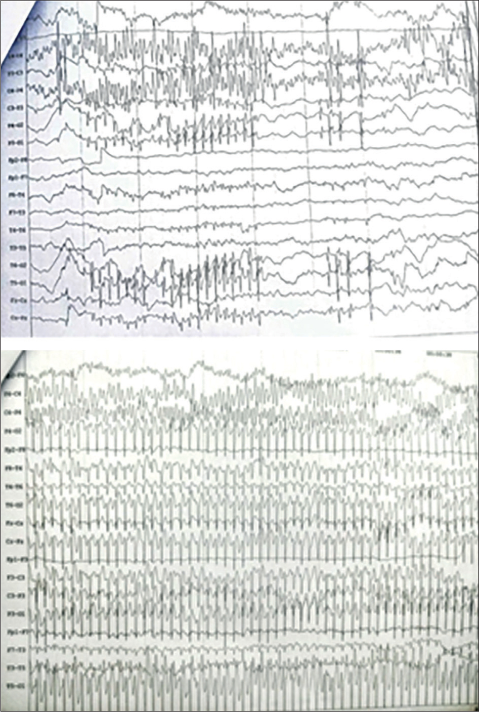
Figure 3.
EEG demonstrating bihemispheric foci with a tendency for generalization.
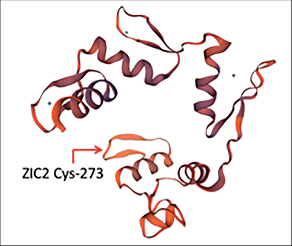
Figure 4.
Molecular model of ZIC2 protein indicating the position of Cys-273.
Table 1.
| GENE | VARIANT | ZYGOSITY | VARIANT CLASSIFICATION |
|---|---|---|---|
| ZIC2 | c.818G>A (p.Cys273Tyr) | heterozygous | Likely Pathogenic |
| ZIC2 | c.1439C>T (p.Ser480Leu) | heterozygous | Uncertain Significance |
| CTNNB1 | c.1657A>G (p.Met553Val) | heterozygous | Uncertain Significance |
| PCLO | c.5293C>T (p.Pro1765Ser) | heterozygous | Uncertain Significance |
| PEX5 | c.1814G>A (p.Ser605Asn) | heterozygous | Uncertain Significance |
| PTPN23 | c.1603C>G (p.Gin535Glu) | heterozygous | Uncertain Significance |