
Table 1
Patient details such as age, gender, tumor locations, cell types and EGFR gene mutation status are shown.
| Sex | Mean Age | Tumor Location (%) | Tumor Cell Type (%) | Status of Analyzed EGFR Gene Mutations Exons (codons) | EGFR Expression | |||
|---|---|---|---|---|---|---|---|---|
| 18 (719) | 19 (deletion) | 20 (768; 790) | 21 (858; 861) | |||||
| F | 57.2±5.6 | duodenum (21.5) | spindle cell (100.0) | wild type | wild type | wild type | wild type | negative |
| ileum (10.5) | epithelioid (100.0) | wild type | wild type | wild type | wild type | negative | ||
| small intestine (26.3); | epithelioid (20.0); mixed (60.0); spindle cell (20.0) | wild type | wild type | wild type | wild type | negative | ||
| stomach (38.8) | spindle cell (57.0); mixed (14.2); epithelioid (28.8) | wild type | wild type | wild type | wild type | negative | ||
| transverse colon (5.2) | mixed (100.0) | wild type | wild type | wild type | wild type | negative | ||
| M | 62.6±10.5 | colon (9.5) | spindle cell (100.0) | wild type | wild type | wild type | wild type | negative |
| descending colon (4.7) | mixed (100.0) | wild type | wild type | wild type | wild type | negative | ||
| duodenum (4.7) | spindle cell (100.0) | wild type | wild type | wild type | wild type | negative | ||
| ileum (4.7) | mixed (100.0) | wild type | wild type | wild type | wild type | negative | ||
| jejenum (4.7) | spindle cell (100.0) | wild type | wild type | wild type | wild type | negative | ||
| small intestine (19.0) | spindle cell (25.0); mixed (75.0) | wild type | wild type | wild type | wild type | negative | ||
| Stomach (52.4) | spindle cell (54.5); mixed (36.3); epithelioid (9.2) | wild type | wild type | wild type | wild type | negative | ||
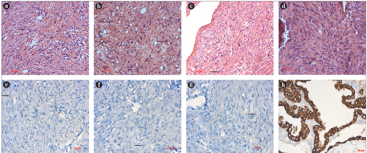
Figure 1
Images a-d show immunohistochemical staining of GIST samples with spindle-shaped tumor cells, negative for EGFR expression. Images e-f indicate hematoxylin and eosin staining of GIST samples with spindle-shaped tumor cells. Image g represents the positive control, expressing EGFR. All images were taken in 40× magnification. Image h represents the control EGFR sample from a chorionic villus sample.