
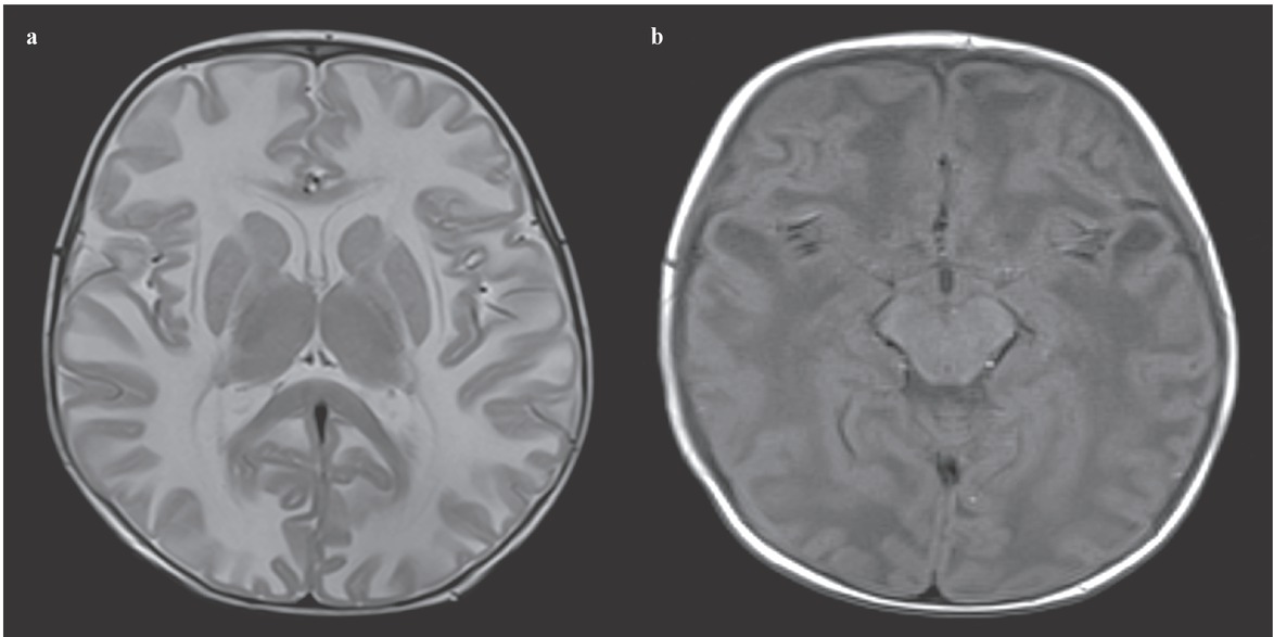
Figure 1
(a) Axial T2 weighted MRI shows bilateral, diffuse, symmetric hyperintense lesions in the cerebral white matter. (b) Axial T1 weighted MRI shows bilateral temporal cysts.
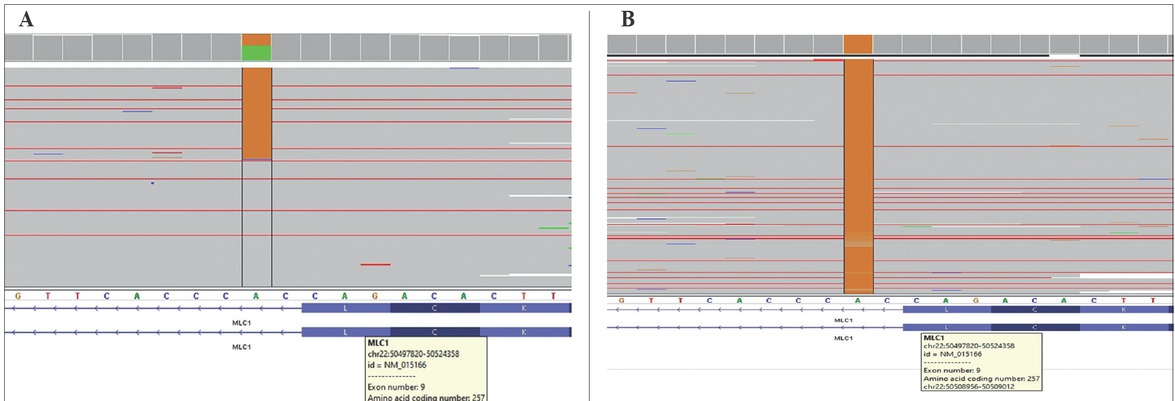
Figure 2
(A) Integrative Genomics Viewer (IGV) sequence data of the parents showed heterozygous c.768+ 2T>C mutation. (B) The IGV sequence data of the patient showed homozygous c.768+2T>C mutation.
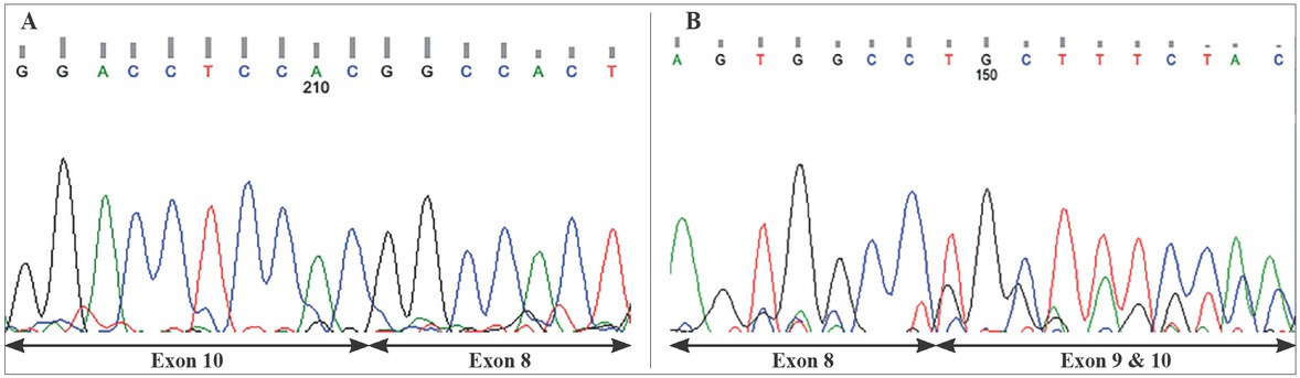
Figure 3
(A) Exon 9 was not found in cDNA (cDNA sequence of the patient, reverse strand). (B) The parents had two strands for the heterozygous mutation. Exons 9 and 10 were overlapping in cDNA (cDNA sequence of the parents, forward strand).