The demand for aesthetically acceptable alternatives to traditional fixed orthodontic appliances has increased, as more adult patients seek treatment.1 Kesling’s positioner, described in 1945, was a precursor to aligner therapy, and subsequently thermoformed splints in the form of retainers have been used to subtly correct the positions of teeth.2 Since the thermoformed splints required significant manufacturing effort and resulted in limited tooth movement, Sheridan expanded the application of vacuum-formed aligners made on casts by removing and grinding portions of the working model, creating windows, deforming the material with specific pliers, arranging composite attachments on the teeth, and adding elastic traction to attachments.2 Subsequently, in 1997, Align Technology, Inc. (Santa Clara, CA, USA) introduced Invisalign®, which applies a sequence of plastic aligners to move teeth following customisation and mass-production to produce personalised appliances.3 Invisalign uses 0.75 mm-thick polyurethane material to manufacture aligners that are worn for one to two weeks before replacement by the next appliance in the series.4 Custom aligners are made using stereolithographic technology from intraoral digital images obtained by scanning in the dental office or, alternatively, from an impression. Maintaining patient compliance is essential for optimal results as patients are required to wear their aligners for at least 22 hr daily.
The expansion of a constricted arch can alleviate crowding, increase the arch length, provide more space for alignment, relieve dentoalveolar posterior crossbites, or enhance a smile’s transverse dimension.5 Using clear aligners, dentoalveolar expansion is a potential alternative to interproximal tooth reduction6,7 although studies on the effectiveness of aligners used for arch expansion are few. The current review aims to identify the best available scientific data on the efficacy of arch expansion by using the Invisalign appliance through a systematic search of the literature.
The present systematic review was registered at the National Institute for Health Research Prospero International Prospective Register of Systematic Reviews (Registration number: CRD42023475837). The research protocol was designed according to the PRISMA (Preferred Reporting Items for Systematic Review and Meta-Analyses) guidelines of 2020.
A) Inclusion Criteria:
1) Participants: Adult patients in the permanent dentition.
2) Intervention: Dentoalveolar expansion using clear aligner therapy.
3) Comparison: Studies evaluating arch width changes before and after clear aligner treatment.
4) Outcome:
i) Primary outcome: Efficiency of arch expansion.
ii) Secondary outcome: Accuracy and Predictability of maxillary arch expansion.
5) Study design: Prospective and retrospective studies in the English language.
B) Exclusion Criteria: In vitro studies, animal studies, case reports, review articles, opinion articles, and letters to editors.
Information Sources, Search Strategy, and Selection Process
A comprehensive search was conducted using electronic databases and supplemented by a manual search, to identify all relevant studies associated with arch expansion as a result of clear aligner therapy. Three electronic databases: PubMed, Google Scholar, and the Cochrane library were searched using the key words “(clear aligner OR permanent dentition) AND (maxillary arch expansion) AND (arch width changes) AND (efficiency OR predictability of maxillary arch expansion)”. A hand search of prominent orthodontic journals was also undertaken while unpublished literature was searched on ClinicalTrials.gov.in. In addition, the reference lists of relevant studies, meta-analyses, systematic reviews, and other review articles were screened for potential inclusion.
The search strategy was developed using relevant Medical Subject Headings (MeSH) terms, keywords, and Boolean operators “AND” and “OR” combinations. The following search terms and their combinations were applied in the search strategy: “(clear aligner OR maxillary arch expansion OR Invisalign) AND (OR efficacy OR predictability of maxillary arch expansion)” The search strategy was adapted to the specific syntax and subject headings of each electronic database.
The selection process followed the Preferred Reporting Items for Systematic Reviews and Meta-Analyses (PRISMA) guidelines. Two reviewers independently screened the titles and abstracts for all relevant articles to determine their eligibility for inclusion. Full-text articles of potentially eligible studies were assessed independently by the two reviewers. Any discrepancies between the reviewers were resolved through discussion or consultation with a third reviewer.
Following the identification of relevant studies, data were extracted using a pre-designed data recording form. The items extracted included the study design, the name of the author, the year of publication, age and gender of the participants, the description of the participants and grouping, the type, and frequency of the intervention, diagnostic methods, observation, and outcomes.
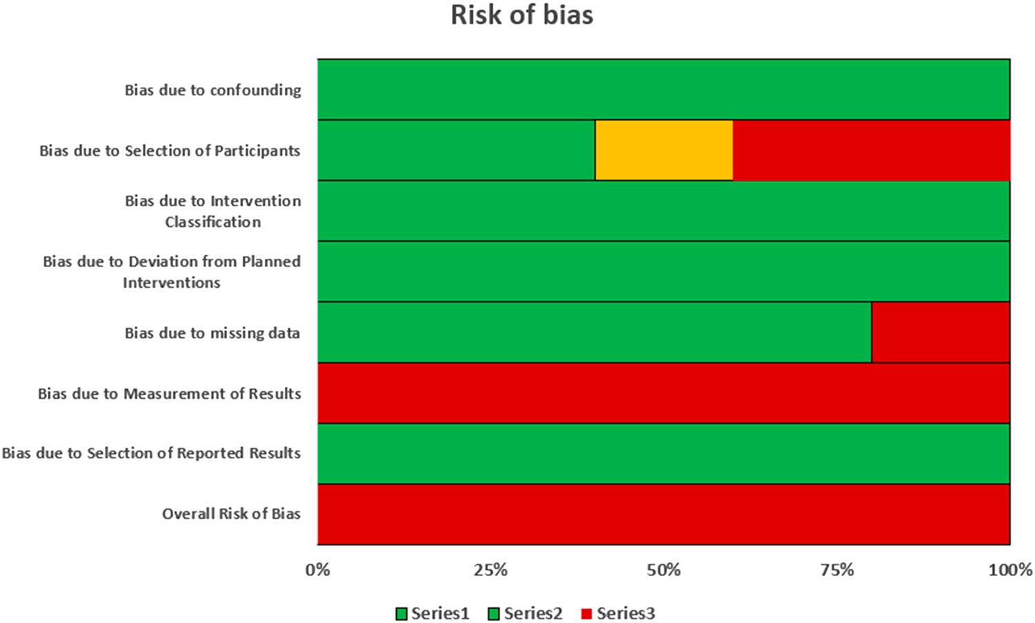
A risk of bias assessment for a non-RCT study was conducted using the ROBINS-I tool and following the recommended approach. The two-part tool was used to address the seven specific bias domains due to confounding, the bias of participant selection, the bias of intervention classification, the bias due to deviations from intended interventions, the bias due to missing data, the bias in the measurement of outcomes, and the bias in the selection of the reported result. The signalling questions were broadly factual in nature and aimed to facilitate judgements about the risk of bias. The categories for the risk of bias judgements were “Low risk”, “Moderate risk”, “Serious risk” and “Critical risk” of bias.
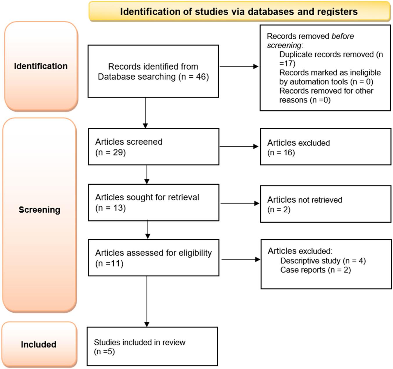
The electronic screening of PubMed and Google Scholar identified 46 articles. After adjusting for duplicates, 29 articles were assessed for inclusion in the present study. The majority were later excluded because of irrelevant titles or abstracts, leaving 11 articles. After excluding four descriptive studies and two case reports, 5 original articles remained for analysis. The PRISMA flowchart of the electronic database search is presented in Figure 1.

Prisma flow diagram of study selection.
Five human studies involving a total population of 254 healthy participants were assessed. Of the five studies, three were prospective and two were retrospective in nature. The demographic details of all of the participants are shown in Table I. Table II lists the parameters examined in each study, as well as the records gathered, and the techniques of collection applied in the data analysis. The results and corresponding conclusions from each study are displayed in Table III.
Demographic details
| Sr. No. | Study ID | Study design | Objectives | Number of patients/sex | Mean age |
| 1. | Jean-Phillippe Houle et al. June, 2017 Angle Orthodontist. | Retrospective study | To investigate the predictability of arch expansion using Invisalign | N = 64 | Mean = 31.2 years Range = 18-61 years |
| 2. | Zhou and Guo August, 2020 | Prospective study | To quantify the efficiency of arch expansion using the Invisalign system in patients, To investigate the movement patterns by comparing actual expansion outcomes of crown and root with virtual planned expansion in ClinCheck software, To ascertain whether the preset expansion amount and initial molar torque correlated with the efficiency of bodily expansion movement. | N = 20 Male = 5 Female = 15 (Chinese adults) | Mean = 28.5 ± 6.3 years Range = 20-45 years |
| 3. | Ignacio Morales-Burruezo et al. December, 2020 PLOS ONE | Retrospective study | To determine the efficacy of the Invisalign system for arch expansion, and to assess the predictability of the measurements planned by Clincheck software for the use of the transparent aligners at the end of the first treatment phase | N = 114 | Range = 18-75 years |
| 4. | Roberta Lione et al. February, 2021 Angle Orthodontist | Prospective study | To evaluate tooth movements during maxillary arch expansion with clear aligner treatment | N = 28 Male = 12 Female = 16 | Mean = 31.9 ± 5.4 years |
| 5. | Gabriella Galluccio et al. March, 2023 International Journal of Environmental Research and Public Health | Prospective study | To evaluate the efficacy and accuracy of maxillary arch transverse expansion using the Invisalign clear aligner system without auxiliaries other than Invisalign attachments. | N = 28 | Mean = 17 ± 3.2 years Range = 13-25 years |
| Sr. No. | Study ID | Groups | Data Collection | Records taken | Parameters measured | Additional parameters |
|---|---|---|---|---|---|---|
| 1. | Jean-Phillippe Houle et al. June, 2017 Angle Orthodontist. |
| Interdental width of both arches
| (For both arches)
| ||
| 2. | Zhou and Guo August, 2020 | According to unilateral expansion for each first molar-
| Pre-expansion (T0) and post-expansion (immediately after finishing N+2 aligner) (T1) records collected, consisting of the following:
| Interdental width of maxillary arches
| For maxillary arch
| — |
| 3. | Ignacio Morales-Burruezo et al. December, 2020 PLOS ONE | According to degree of complexity of transverse malocclusion-
|
| Measurements were taken from occlusal images captured at T1, T2, and T3-
|
| |
| 4. | Roberta Lione et al. February, 2021 Angle Orthodontist |
|
| — | — | |
| 5. | Gabriella Galluccio et al. March, 2023 International Journal of Environmental Research and Public Health | An intraoral scan were performed with the Itero Flex® scanner and final position of ClinCheck® representation (TC) collected. Models collected were-
| The following transverse linear measurements were carried out on the upper arch for each T0 and T1 model and for the ClinCheck® model (TC)
|
| Clinical accuracy (%) was achieved for all measurements, using the equation [(expansion obtained/planned expansion) × 100]. |
| No. | Study ID | Outcome | Conclusion |
|---|---|---|---|
| 1. | Jean-Phillippe Houle et al. June, 2017 Angle Orthodontist. |
|
|
| 2. | Zhou and Guo August, 2020 |
|
|
| 3. | Ignacio Morales-Burruezo et al. December, 2020 PLOS ONE |
|
|
| 4. | Roberta Lione et al. February, 2021 Angle Orthodontist |
|
|
| 5. | Gabriella Galluccio et al. March, 2023 International Journal of Environmental Research and Public Health |
|
|
The risk of bias assessment for non-RCT studies is summarised in Table IV. There was a significant risk of bias in every reviewed study, with patient selection and the lack of a blinded assessment being the most frequent sources. None of the trials addressed the result of the assessor’s blinding. A significant risk of bias was indicated by two investigations (Houle et al. and Morales-Burruezo)6,7 because of the wide age range of the patients. Poor compliance was cited as a reason for patient attrition in a study by Lione et al.,8 thereby indicating a significant risk of bias (Figure 2).

Risk of bias graph for the included studies.
| Study | Pre-intervention | Intervention | Post-intervention | |||||
|---|---|---|---|---|---|---|---|---|
| Risk of bias of confounding | Risk of bias in the selection of participants | Risk of bias in the intervention classification | Risk of bias as a result of deviation from planned intervention | Risk of bias as a result of missing data | Risk of bias in the measurement of results | Risk of bias in the selection of reported results | General Judgment of Risk of Bias | |
| Houle et al 2017 | Low | Serious | Low | Low | Low | Serious | Low | Serious |
| Zhou and Gou 2020 | Low | Moderate | Low | Low | Low | Serious | Low | Serious |
| Morales-Burruezo et al 2020 | Low | Serious | Low | Low | Low | Serious | Low | Serious |
| Lione et al 2021 | Low | Low | Low | Low | Serious | Serious | Low | Serious |
| Galluccio et al 2023 | Low | Low | Low | Low | Low | Serious | Low | Serious |
Comparison of change in the inter-canine width before and after aligner treatment (Figure 3).

Comparison of the change in inter-canine width before and after orthodontic treatment.
A meta-analysis was performed on three studies that qualified and provided the required data outcome for a quantitative analysis depicted as a forest plot. Because heterogeneity was more than 50% (I2 = 0%) following the conducted meta-analysis, a fixed effect model was applied. The change in inter-canine width after aligner treatment was greater than the inter-canine width before treatment (SMD: -0.80; 95% CI = -1.02 to -0.58; Z value = 7.06), and the difference was statistically significant (p < 0.00001).
Comparison of the inter-first premolar width before and after aligner treatment (Figure 4).

Comparison of the inter-first premolar width before and after orthodontic treatment.
A meta-analysis was performed on three studies that qualified with the required dataset that could be quantitatively analysed and depicted as a forest plot. Heterogeneity was more than 50% (I2 = 0%) and so a fixed effect model was applied. The change in inter-first premolar width after the orthodontic treatment was greater than the inter-first premolar width before treatment (SMD: -1.13; 95% CI = -1.36 to -0.90; Z value = 9.62), and the difference was statistically significant (p < 0.00001).
Comparison of the inter-second premolar width before and after aligner treatment (Figure 5).

Comparison of the inter-second premolar width before and after orthodontic treatment.
A meta-analysis was performed on three studies that contained the required data that could be quantitatively analysed and depicted as a forest plot. Heterogeneity was more than 50% (I2 = 0%) and so a fixed effect model was applied. The change in inter-second premolar width after the aligner treatment was greater than the inter-second premolar width before treatment (SMD: -1.07; 95% CI = -1.30 to -0.84; Z value = 9.18), and the difference was statistically significant (p < 0.00001).
Comparison of the inter-molar width before and after aligner treatment (Figure 6).

Comparison of the inter-molar width before and after orthodontic treatment.
A meta-analysis was performed on four comparisons from three studies that contained the required dataset for quantitative analysis and depicted as a forest plot. Heterogeneity was more than 50% (I2 = 0%) and so a fixed effect model was applied. The change in inter-molar width after the aligner treatment was greater than the inter-molar width before treatment (SMD: -0.72; 95% CI = -0.93 to -0.52; Z value = 6.69), and the difference was statistically significant (p < 0.00001).
Comparison of the change in inter-canine width after aligner treatment with the change predicted by the ClinCheck software (Figure 7).

Comparison of the change in intercanine width after orthodontic treatment with the change predicted by the ClinCheck software.
A meta-analysis was performed on five studies that provided the required dataset for quantitative analysis and depicted as a forest plot. Heterogeneity was more than 50% (I2 = 71%) and so a random effect model was applied. The change in inter-canine width after aligner treatment was lower than that predicted by the ClinCheck software (SMD: 0.57; 95% CI = 0.20 to 0.93; Z value = 3.06), and the difference was statistically significant (p = 0.002).
Comparison of the change in inter-first premolar width after aligner treatment with the change predicted by the ClinCheck software (Figure 8).

Comparison of the change in inter-first premolar width after orthodontic treatment with the change predicted by the ClinCheck software.
A meta-analysis was performed on five studies that provided the required dataset for quantitative analysis and depicted as a forest plot. Heterogeneity was more than 50% (I2 = 90%) and so a random effect model was applied. The change in inter-first premolar width after orthodontic treatment was lower than that predicted by the ClinCheck software (SMD: 0.57; 95% CI = -0.06 to 1.20; Z value = 1.78) but the difference was statistically non-significant (p = 0.008).
Comparison of the change in inter-second premolar width after aligner treatment with the change predicted by the ClinCheck software (Figure 9).

Comparison of the change in inter-second premolar width after orthodontic treatment with the change predicted by the ClinCheck software.
A meta-analysis was performed on five studies that provided the required dataset for quantitative analysis and depicted as a forest plot. Heterogeneity was more than 50% (I2 = 85%) and so a random effect model was applied. The change in inter-second premolar width after orthodontic treatment was lower than that predicted by the ClinCheck software (SMD: 0.51; 95% CI = 0.02 to 1.01; Z value = 2.03) and the difference was statistically significant (p = 0.04).
Comparison of the change in inter-molar width after aligner treatment with the change predicted by the ClinCheck software (Figure 10).

Comparison of the change in inter molar width after orthodontic treatment with the change predicted by the ClinCheck software.
A meta-analysis was performed on five studies that provided the required dataset for quantitative analysis and depicted as a forest plot. Heterogeneity was more than 50% (I2 = 97%) and so a random effect model was applied. The change in inter-molar width after orthodontic treatment was lower than that predicted by the ClinCheck software (SMD: 0.93; 95% CI = -0.22 to 2.08; Z value = 1.59), but the difference was statistically non-significant (p = 0.11).
The present systematic review summarises evidence from prospective and retrospective studies which evaluated the effectiveness of maxillary arch expansion by clear aligners in adult patients. Invisalign aligners have been shown to be effective in treating complicated malocclusions, by buccal expansion of approximately 2 to 4 mm achieved either to reduce crowding or to alter arch form.6 It is crucial that clinicians appreciate the effectiveness and consequences of Invisalign aligner-based arch expansion therapy on skeletal and dental components. Interdental width has previously been solely assessed for tooth crowns using 3D digital model measurements; however, the buccal movement of tooth roots and skeletal effects have not been assessed. The five studies included in the present review fulfilled the research criteria and evaluated the effectiveness and efficiency of maxillary arch expansion by clear aligners in adult patients, (mean difference between pre-treatment arch width and arch width after arch expansion) and the predictability of the planned arch expansion (mean difference between predicted and actual achieved arch expansion). In each of the five reports, pre-treatment and post-treatment digital models (.stl files) generated from an iTero scan were obtained. The models were used to compare the final generated virtual model in relation to the movements seen in the post-treatment model, by using the ClinCheck software.
In addition to the treatment efficiency and predictability of the planned arch expansion, molar inclination was evaluated by Lione et al.8 and Morales-Burruezo et al.9 Interdental gingival measurements were also taken into account by Houle et al.6 and Galluccio et al.,10 and molar rotation was additionally evaluated by Morales-Burruezo et al.9 The results of Zhou and Guo5 demonstrated that Invisalign aligners can achieve arch expansion, with efficiencies of 79.75 ± 15.23% at the canine cusp tip, 76.1 ± 18.32% at the first premolar cusp tip, 73.27 ± 19.91% at the second premolar cusp tip, and 68.31 ± 24.41% at the first molar cusp tip. The expansion efficiencies were marginally less than those reported by Houle et al.6 who indicated that the canine, first premolar crown, second premolar, and the first molar had expansion efficiencies of 88.7%, 84.7%, 81%, and 76.6%, respectively. The accuracy of tooth movement for labial expansion of the anterior teeth was reported to be 40.5% by Kravitz et al.11
The level of anticipated expansion determined the disparity in predictability between the groups. According to a Spearman correlation (P = 0.05), Zhou and Guo5 reported that the present expansion quantity and physiological expansion efficiency had a negative link. Houle et al. found that the overall accuracy of transverse changes in the upper arch was 72.8%, with 82.9% accuracy at the cusp tips and 62.7% accuracy at the gingival margins. With an accuracy of 88.7%, the canine cusp tip was the most accurate forecast which means that 0.22 mm of the 1.92 mm desired expansion was not attained. The accuracy was 52.9% at the gingival margin of the first molar.
Gingival margin measurements revealed that the lower canines had the most prediction error (62% accuracy), followed by the molars (85.5%), the first premolar (88.4%), and the molars at 70.7% overall. Since the amount of change required in the lower arch is typically less than in the upper arch, this could account for the higher outcome.6 The predictability data from Morales-Burruezo et al.9 suggested that the behaviour of aligners is comparable to that of conventional orthodontic arch wires, when considering deflection and inter-tooth distance. However, the system predictability results were not comparable.
Even though the identified studies were closely matched for sample size, there was no consideration of aligners made from the SmartTrack material. Furthermore, all of the patients received additional aligners (ranging from one to five phases of refinement treatment), and the system predictability results were not comparable.12 Despite this, the study examined 64 patients (20 of whom had at least one tooth in crossbite) during the initial phase of therapy without the need for additional aligners, even though these were made of EX30 material.13
In summary, a steady decrease in the accuracy of the rate of expansion from the anterior to the posterior segment was observed in the five studies. This may be attributed to differences in root anatomy and cortical bone thickness, a higher occlusal load, greater soft tissue resistance in the posterior region, and a decline in mechanical efficiency from the anterior to the posterior segments. The efficacy of expansive movement was substantially inversely related to the preset expansion quantity and the initial torque of the maxillary first molar.
In addition, the change in inter-canine, inter-first premolar, inter-second premolar, and inter-molar width after the aligner treatment was greater than the inter-canine, inter-first premolar, inter-second premolar, and inter-molar width before orthodontic treatment. The change in inter-canine, inter-first premolar, inter-second premolar, and inter-molar width after orthodontic treatment was less than that predicted by the ClinCheck software. At the end of an aligner treatment sequence, appliance efficiency may be slightly higher due to hysteresis, which is the lag between the expected and obtained results (the difference between the expected and the actual obtained expansion) of the Invisalign appliances. This lag is mostly related to energy loss that occurs during treatment because of the material’s viscoelastic properties.
The present systematic review revealed that clear aligner treatment has potential benefits for orthodontic patients requiring arch expansion. To enable better and more effective aligner therapy, further research is needed to address issues as the average age of patients receiving treatment is steadily rising.
The effectiveness and predictability of maxillary arch expansion in adult patients receiving clear aligner therapy were examined in the present systematic review. Retrospective and prospective studies were considered but case reports were excluded, and non-English language material was eliminated using a language filter. Due to their recency, heterogeneity, and non-comparable diagnostic techniques, all studies carried a significant risk of bias. The review assessed the outcome efficacy in spite of these drawbacks.
An area of focus has been the effectiveness of maxillary arch expansion in adult patients receiving clear aligner therapy. The hysteresis (lag) of the Invisalign devices may cause appliance efficiency to be slightly higher at the completion of an aligner treatment sequence. Over the course of treatment, the maxillary arch’s expansion rate declined progressively in the canine, premolar, and posterior regions, with the first and second premolars exhibiting the largest net expansion. When planning aligner arch expansion, a thorough assessment and overcorrection are crucial because the arch expansion efficiency is adversely correlated with the starting torque and level of required expansion. This strategy can assist in reducing the frequency of mid-course adjustments and revisions during the course of care.