
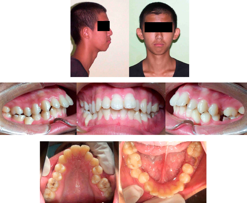
Figure 1.
Pre-treatment facial and intraoral photographs.
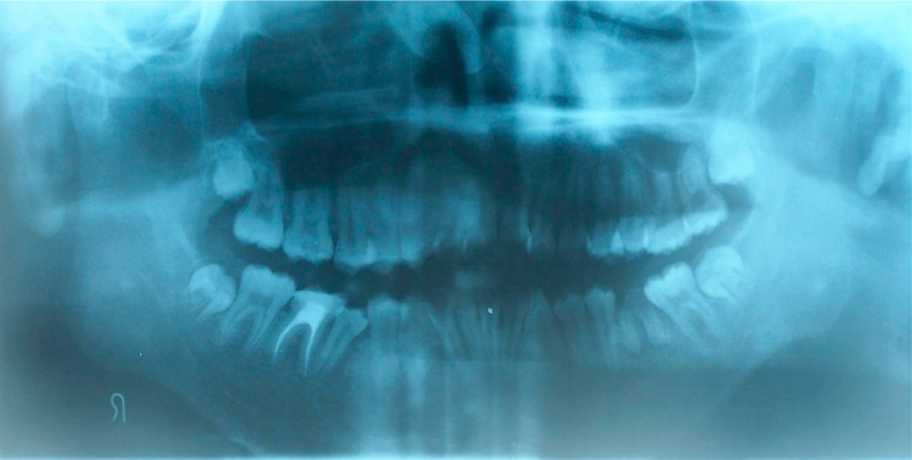
Figure 2.
Pretreatment radiographs: Panoramic radiograph.
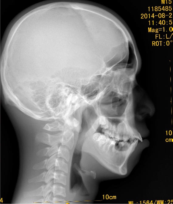
Figure 3.
Pretreatment radiographs: Lateral cephalogram.
Table I.
Cephalometric analysis
| Chinese norm | Pre-treatment | Post-treatment | |
|---|---|---|---|
| SNA (°) | 82.8 ± 4.0 | 80.6 | 80.1 |
| SNB (°) | 80.1 ± 3.9 | 77.2 | 77.6 |
| ANB (°) | 2.7 ± 2.0 | 3.4 | 2.5 |
| NA-PA (°) | 6.0 ± 4.4 | 7.5 | 4.7 |
| FMA (°) | 31.3 ± 5.0 | 31.7 | 31.9 |
| SN-MP (°) | 31.1 ± 5.6 | 37.8 | 38.3 |
| U1-SN (°) | 105.7 ± 6.3 | 118.7 | 107.4 |
| U1-NA (mm) | 5.1 ± 2.4 | 11.2 | 3.0 |
| L1-MP (°) | 92.6 ± 7.0 | 91.4 | 85.5 |
| L1-NB (mm) | 6.7 ± 2.1 | 10.5 | 4.0 |
| Interincisal angle (°) | 125.4 ± 7.9 | 111.7 | 127.5 |
| Y-axis angle (°) | 65.8 ± 4.2 | 68.6 | 64.3 |
| Overbite (mm) | 0-3.0 | 3.6 | 0.8 |
| Overjet (mm) | 0-3.0 | 6.0 | 1.9 |
| Upper lip to E-line (mm) | -1.0 ± 2.0 | 4.0 | -1.4 |
| Lower lip to E-line (mm) | 0.0 ± 2.0 | 7.8 | -0.2 |
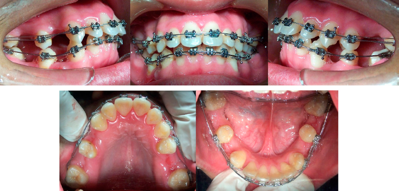
Figure 4.
Intraoral photographs at 1 month.
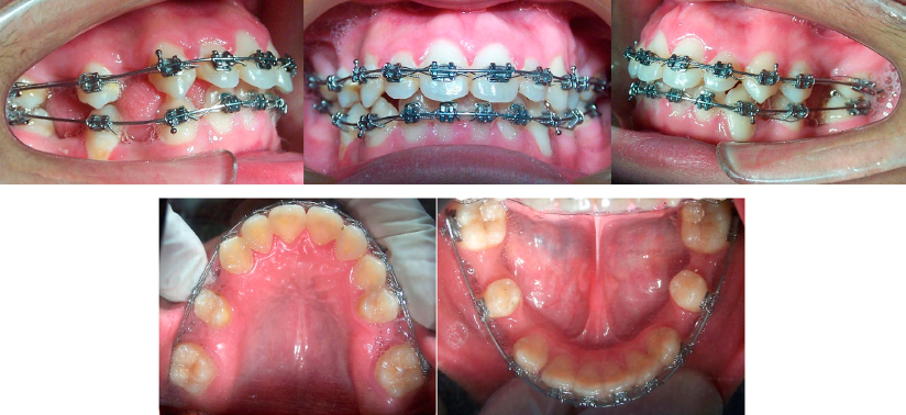
Figure 5.
Intraoral photographs at 8 months.
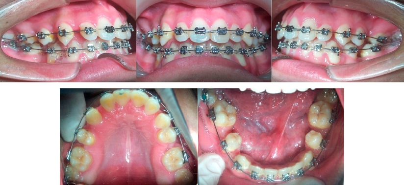
Figure 6.
Intraoral photographs at 21 months.
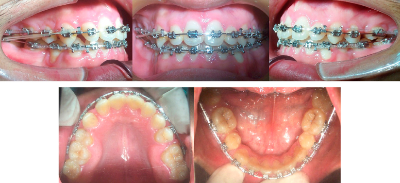
Figure 7.
Intraoral photographs at 35 months.
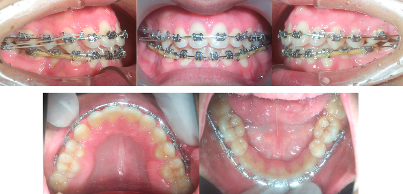
Figure 8.
Intraoral photographs at 44 months.
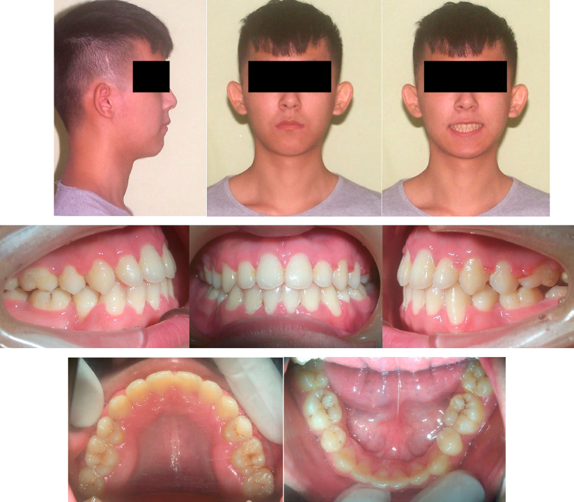
Figure 9.
Post-treatment facial and intraoral photographs.
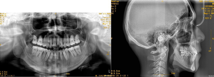
Figure 10.
Post-treatment radiographs: (A) Panoramic radiograph, (B) Lateral cephalogram.
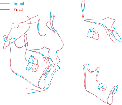
Figure 11.
Lateral cephalogram and superimposition tracings.
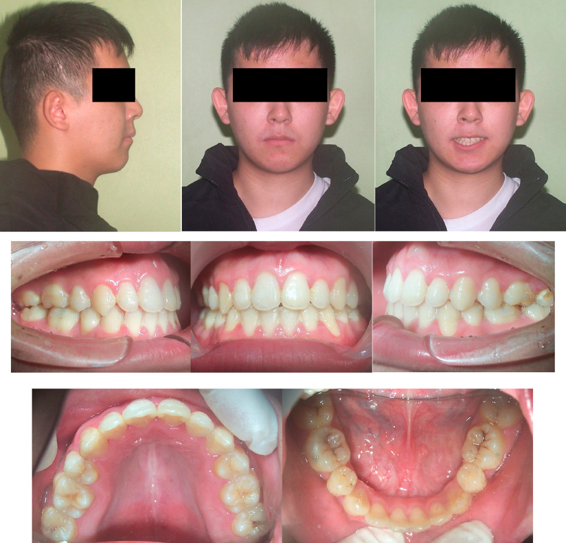
Figure 12.
Five-year retention facial and intraoral photographs.