
Table 1.
Medium conductivity (mS) depending on time of ozonation and time after ozonation
| Ozonation time (s) | Conductivity measurement after ozonation (mS) Average of 5 measurements, deviation ± 1 mS | ||
|---|---|---|---|
| Immediately after ozonation | After 24 h of keeping medium in 4°C | After 48 h of keeping medium in 4°C | |
| 6 | 12 | 11 | 9 |
| 12 | 15 | 12 | 10 |
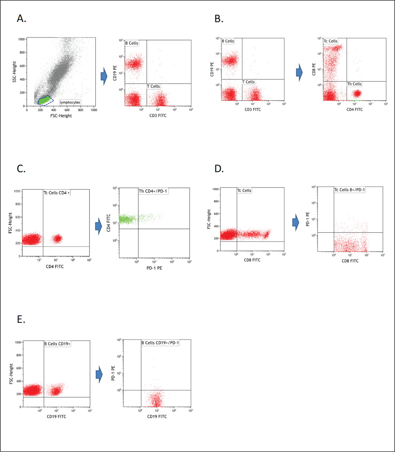
Fig 1.
An example of the flow cytometry analysis of antigens expression on T and B lymphocytes. (A) Gating strategy for lymphocytes as well as their subpopulations, B lymphocytes (CD19+) and T lymphocytes (CD3+); (B) Gating strategy for Tc (CD8+) and Th (CD4+) lymphocytes among T cell subpopulations (CD3+); (C) Strategy for gating PD-1 expression among the Th cell population; (D) Gating strategy for PD-1 expression among the Tc cell population; (E) Gating strategy for PD-1 expression among the B cell population. PD-1, programmed cell death protein 1.
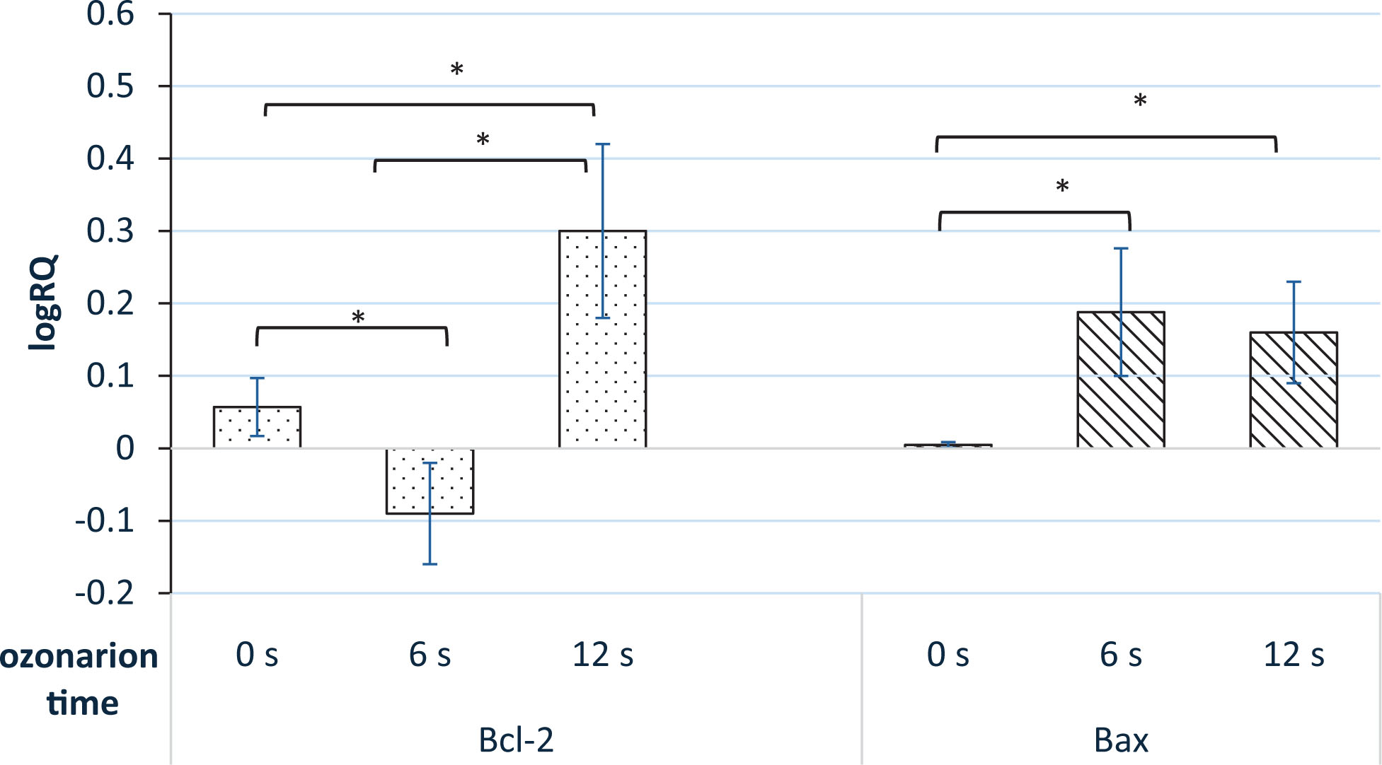
Fig 2.
Average relative expression of Bcl-2 and Bax genes (logRQ) in dental pulp cells cultures depending on the ozonation time compared to cells without ozone treatment. The cells not subjected to ozonation (ozone dose 0 s.) or exposed to ozone for 6 s or 12 s were analyzed for the expression of Bax and Bcl-2. Data are representative of six independent experiments, each repeated in duplicate ± SD; *Indicates significant difference between two indicated groups (p < 0.05, ANOVA test).
Table 2.
Bax/Bcl-2 expression ratio in response to ozonation of dental pulp cells
| Ozonation time (s) | Bax mean (logRQ) | Bcl-2 mean (logRQ) | Bax/Bcl-2 ratio | p-value |
|---|---|---|---|---|
| 0 | 0.0048 | 0.0573 | 0 | 0.199 |
| 6 | 0.1883 | −0.0903 | −2 | 0.0011* |
| 12 | 0.1611 | 0.3 | 0.5 | 0.0005*,** |
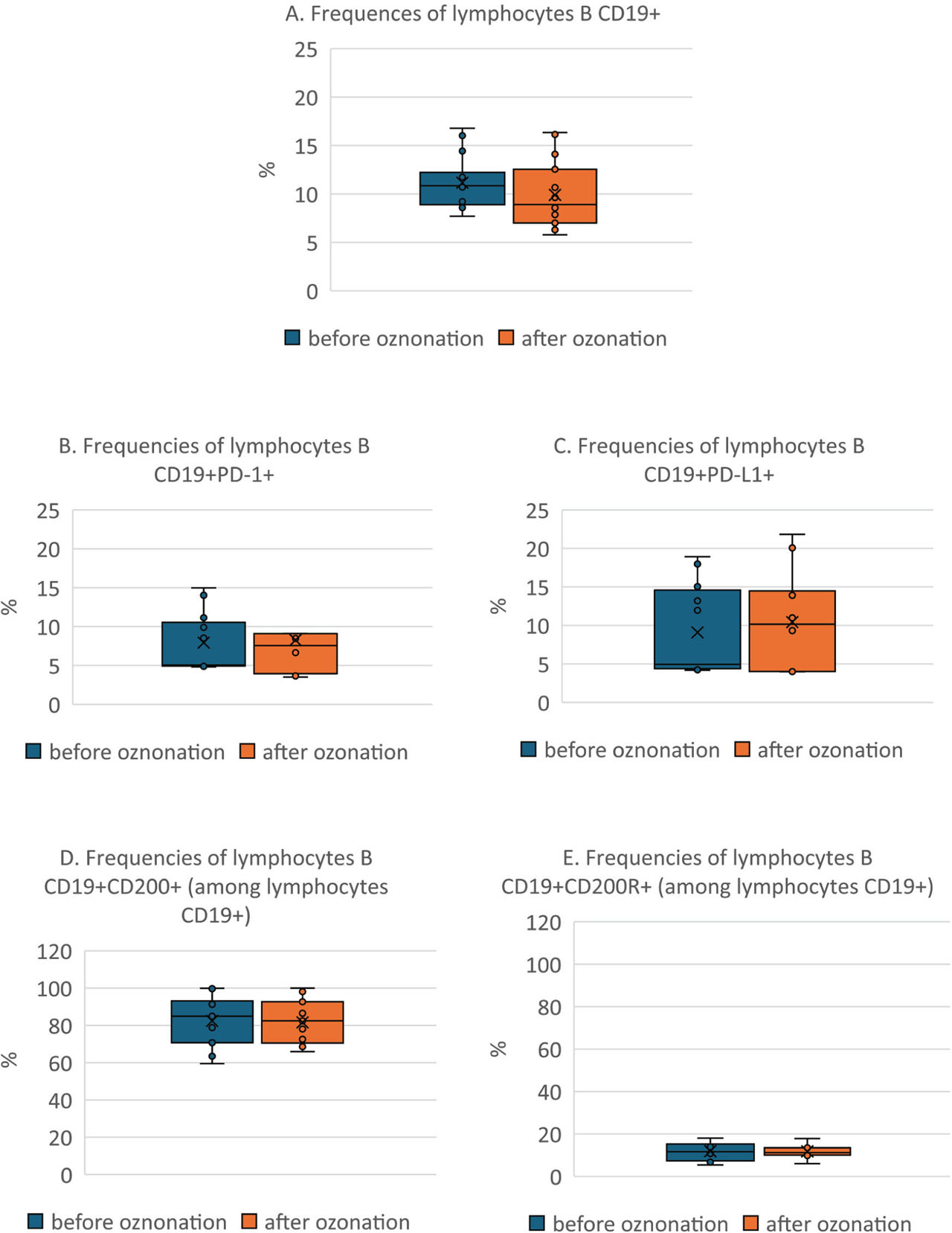
Fig 3.
Differences in B lymphocyte measurements before and after ozonation. The percentages of CD19+ B lymphocytes (A) expressing PD-1 (B) and PD-L1 (C) molecules in the studied cell populations isolated from dental pulp of healthy donors (n=15) before and after ozonation. CD19+CD200 expression (D) and CD19+CD200R expression in B lymphocytes (E) before and after ozonation. Dots represent the cases (n = 15), (—) median, (×) mean, whiskers indicate quartiles. PD-1, programmed cell death protein 1; PD-L1, programmed death-ligand 1.
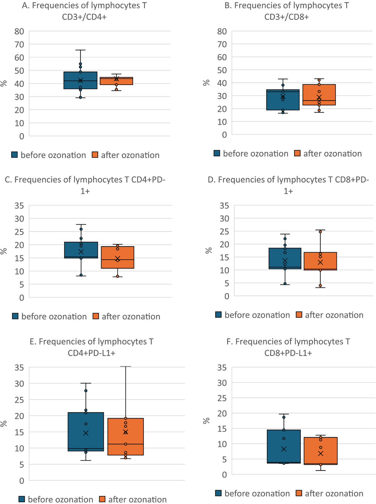
Fig 4.
Differences in T lymphocyte measurements before and after ozonation. The percentages of CD4+ (A) and CD8+ (B) T lymphocytes expressing PD-1 (B, E) and PD-L1 (C, F) molecules in the studied cell populations isolated from dental pulp of healthy donors (n = 15) before and after ozonation. Dots represent the cases (n = 15), (—) median, (×) mean, whiskers indicate quartiles. PD-1, programmed cell death protein 1; PD-L1, programmed death-ligand 1.