
Table 1
Substances and their concentrations used in experiments
| Name | Concentration (μg/mL) | Description |
|---|---|---|
| Nitrofurantoin* | 167 (0.16 % DMSO) | prophylactic dose (100 mg of nitrofurantoin consumed orally) |
| 330 (0.3 % DMSO) | minimal therapeutic dose (50 mg, 4× daily – 200 mg/day) | |
| 670 (0.6 % DMSO) | maximal therapeutic dose (100 mg, 4× daily – 400 mg/day) | |
| Hydroquinone | 5; 20; 50; 320; 900 | concentrations that are present in the urinary bladder after standard therapeutic application of bearberry extract (15) |
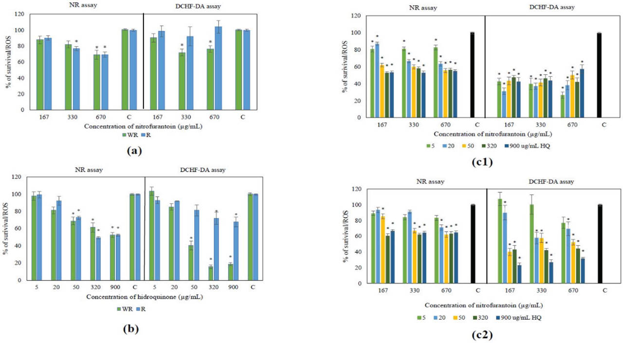
Figure 1
Cytotoxic (neutral red) and antioxidative/prooxidative (DCHF-DA) activity of a) nitrofurantoin, b) hydroquinone, and their mixture in unrecovered (c1) and recovered over 24 h (c2) human urinary bladder cancer T24 cells. * Statistically significant result compared to the control (100 %; one-way ANOVA followed by post hoc Tukey's HSD test; P<0.05)
Table 2
Benchmark dose (BMD) and confidence intervals BMDI (BMDL-BMDU) for hydroquinone plus nitrofurantoin mixtures and results of factorial regression analyses to determine their effect on T24 cell survival and induction of ROS after 8 h exposure
| Treatment | Response | PROAST software# | STATISTICA software* | Activity | ||||||
|---|---|---|---|---|---|---|---|---|---|---|
| BMD | BMDL (μg/mL) | BMDU | Effect | Multiple R (model testing) | P-value | β12 | P-value | |||
| Unrecovered cells | % of survival | 10.64 | 5.76 | 15.2 | yes | 0.64 | <0.01 | +2.21×10⁻⁵ | 0.11 | / |
| Recovered cells (after 24 h) | % of survival | 33.99 | 21.6 | 49 | yes | 0.72 | <0.01 | +9.38×10⁻⁵ | <0.01 | synergistic |
| Unrecovered cells | ROS induction | 16.04 | 13.6 | 21.2 | yes | 0.53 | <0.01 | +0.0001 | <0.01 | synergistic |
| Recovered cells (after 24 h) | ROS induction | 15.4 | 0.532 | 227 | no | 0.62 | <0.01 | −1.55×10⁻⁵ | 0.48 | / |
Table 3
Formation of micronuclei (MN), nucleoplasmic bridges (NPB), and nuclear buds (NB) in T24 cells after treatment with nitrofurantoin (N), hydroquinone (HQ), and their mixture over 2 and 8 h
| Sample | Time | Micronuclei | Nucleoplasmic bridges | Nuclear buds | ||||||||
|---|---|---|---|---|---|---|---|---|---|---|---|---|
| Total (MN)6000 ± SD | Mean (MN)1000 ± SD | Total (BNMN)6000 ± SD | Mean (BNMN)1000 ± SD | Distribution of BNMN cells with | ||||||||
| 1 MN | 2 MN | 3 MN | Mean (NPB)1000 ± SD | Total (NPB)6000 ± SD | Mean (NB)1000 ± SD | Total (NB)6000 ± SD | ||||||
| NC1 | 2 h | 86 | 14.3±0.52 | 78 | 13±1.05 | 71 | 6 | 1 | 1.2±0.75 | 7 | 1.3±0.75 | 8 |
| NC2 | 75 | 12.5±1.03 | 69 | 11.5±0.53 | 63 | 6 | 0 | 1.5±0.84 | 9 | 0.7±0.51 | 4 | |
| N (167 μg/mL) | ↑135e | 22.5±3.99 | ↑127 | 21.2±4.36 | 118 | 7 | 1 | 9±2.53 | ↑54d,e | 5±1.55 | ↑30e | |
| HQ (5 μg/mL) | ↑125e | 20.8±4.02 | ↑110e | 18.3±2.94 | 96 | 13 | 1 | 10.3±1.51 | ↑62d | 5.5±1.52 | ↑33e | |
| HQ (10 μg/mL) | ↑145e | 24.2±2.93 | ↑135 | 22.5±2.26 | 125 | 10 | 0 | 18.5±3.39 | ↑111a,b,c | 6.2±1.47 | ↑37a | |
| MIX 1 | ↑187a,b,c,d | 31.2±5.15 | ↑165a,c | 27.5±4.46 | 141 | 23 | 0 | 14.2±4.79 | ↑85a,b | 8.5±2.2 | ↑51a,b | |
| MIX 2 | / | / | / | / | / | / | / | / | / | / | / | |
| PC | ↑138e | 23 3±3.33 | ↑107e | 17.8±2.21 | 17 | 17 | 4 | 7.3±2.1 | ↑44d,e | 3.5±0.92 | ↑21d | |
| NC1 | 8 h | 72 | 12 1±.09 | 68 | 11.3±1.37 | 6 | 6 | 0 | 1.2±0.41 | 7 | 0.8±0.05 | 5 |
| NC2 | 65 | 10.8±1.47 | 62 | 10.3±1.51 | 2 | 2 | 0 | 1±0.15 | 6 | 0.7±0.03 | 4 | |
| HQ (5 μg/mL) | ↑167d | 27.8±2.99 | ↑143d | 23.8±1.17 | 18 | 18 | 3 | 30.2±2.08 | ↑46a | 5.5±1.05 | ↑33a,c | |
| HQ (10 μg/mL)* | ↑133a,c, * | 22.2±3.65 | ↑112a,c, * | 18.7±2.73 | 13 | 13 | 4 | 19±1.03 | ↑19a, * | 7.3±1.97 | ↑44a,b, * | |
| PC | ↑147d | 24.5±3.99 | ↑133d | 22.2±3.13 | 14 | 14 | 0 | 11.8±3.06 | ↑71b,c | 8.5±1.16 | ↑51b,c | |
NC1 – negative control, non-treated cells; N – nitrofurantoin; HQ – hydroquinone; MIX 1 – mixture of N (167 μg/mL) and HQ (5 μg/mL); MIX 2 – mixture of N (167 μg/mL) and HQ (10 μg/mL); PC – positive control (0.4 μg/mL); NC2 – negative control, cells treated with 0.16 % DMSO used for stock of nitrofurantoin preparation.
Table 4
Cell viability, cytostatic effect, and parameters of cell proliferation in T24 cells after treatment with nitrofurantoin, hydroquinone, and their mixture over 2 and 8 h
| Sample | Time | AP (%) | Cytostatic effect | Parameters of cell proliferation | ||
|---|---|---|---|---|---|---|
| CBPI | % cytostasis | NDI | RI (%) | |||
| NC | 2 h | 0.13 | 1.718 | 0 | 1.726 | 100 |
| N (167 μg/mL) | 0.17 | ↓1.445 | 27.3 | ↓1.447 | 61.94 | |
| N (330 μg/mL) | 0.18 | ↓1.259 | 45.9 | ↓1.261 | 36.94 | |
| HQ (5 μg/mL) | 0.15 | 1.695 | 2.3 | 1.699 | 96.82 | |
| HQ (10 μg/mL) | 0.25 | 1.666 | 5.2 | 1.671 | 92.89 | |
| MIX 1 | 0.13 | ↓1.225 | 49.3 | ↓1.225 | 31.28 | |
| MIX 2 | 0.15 | ↓1.077 | 64.1 | ↓1.077 | 10.68 | |
| NC | 8 h | 0.17 | 1.558 | 0 | 1.561 | 100 |
| N (167 μg/mL) | 0.25 | ↓1.058 | 50.0 | ↓1.059 | 10.29 | |
| HQ (5 μg/mL) | 0.28 | 1.449 | 10.9 | 1.453 | 80.51 | |
| HQ (10 μg/mL) | ↑0.83* | ↓1.229 | 32.9 | ↓1.231 | 40.96 | |
AP – number of cells in apoptosis (%); CBPI – Cytokinesis-block proliferation index; HQ – hydroquinone; MIX 1 – mixture of N (167 μg/mL) and HQ (5 μg/mL); MIX 2 – mixture of N (167 μg/mL) and HQ (10 μg/mL); N – nitrofurantoin; NC – negative control; NDI – nuclear division index; RI – replication index. ↓ significantly lower than NC (P<0.05; chi-squared test).
Table 5
Proteomic analysis results obtained after treatment with nitrofurantoin, hydroquinone, and their mixture over 8 h (fold change cut-off ≥1.5 and ≤0.667)
| Condition | Protein namea | Accession numberb | Alternate IDc | Permutation test (P-value) | Fold changed |
|---|---|---|---|---|---|
| Hydroquinone | Albumin | A0A0C4DGB6 | ALB | < 0.0001 | 0.3 |
| Guanine nucleotide-binding protein G(s) subunit alpha isoforms short | A0A7I2V5R6 | GNAS | 0.001 | 0.4 | |
| Sideroflexin-1 | Q9H9B4 | SFXN1 | 0.004 | 0.5 | |
| THO complex subunit 4 | E9PB61 | ALYREF | < 0.0001 | 0.4 | |
| CD44 antigen | P16070 | CD44 | < 0.0001 | 0.5 | |
| Tropomyosin alpha-3 chain | A0A087WWU8 | TPM3 | 0.003 | 0.5 | |
| Zinc transporter ZIP3 | F5H385 | SLC39A3 | 0.003 | 0.3 | |
| Heterogeneous nuclear ribonucleoprotein U | A0A1W2PPS1 | HNRNPU | 0.002 | 0.5 | |
| Cathepsin B | A0A7P0NGZ6 | CTSB | 0.002 | 0.5 | |
| Succinate–CoA ligase [ADP/GDP–forming] subunit alpha, mitochondrial | P53597 | SUCLG1 | 0.001 | 0.6 | |
| Non-histone chromosomal protein HMG–17 | P05204 | HMGN2 | 0.003 | 0.55 | |
| Procollagen–lysine,2–oxoglutarate 5–dioxygenase 2 | O00469 | PLOD2 | < 0.0001 | 1.5 | |
| Synaptic vesicle membrane protein VAT–1 homolog | Q99536 | VAT1 | 0.003 | 1.7 | |
| Filamin-C OS=Homo sapiens | Q14315 | FLNC | 0.0009 | 1.7 | |
| Mesencephalic astrocyte-derived neurotrophic factor | P55145 | MANF | 0.004 | 1.7 | |
| Tyrosine-protein phosphatase non–receptor type 1 | P18031 | PTPN1 | 0.0003 | 2.3 | |
| Delta–1–pyrroline–5–carboxylate synthase | P54886 | ALDH18A1 | < 0.0001 | 1.9 | |
| UDP–glucose:glycoprotein glucosyltransferase 1 | Q9NYU2 | UGGT1 | < 0.0001 | 2.0 | |
| Histone H1.4 | P10412 | H1–4 | 0.002 | 2.4 | |
| Lysophospholipid acyltransferase 7 | Q96N66 | MBOAT7 | 0.001 | 4.2 | |
| ATP-dependent 6–phosphofructokinase, platelet type | Q01813 | PFKP | 0.001 | 4.4 | |
| LIM domain only protein 7 | F8WD26 | LMO7 | 0.001 | 3.3 | |
| MARCKS–related protein | P49006 | MARCKSL1 | 0.002 | 8.1 | |
| Aminoacyl–tRNA hydrolase | J3KQ48 | PTRH2 | 0.0001 | 5.6 | |
| A–kinase anchor protein 12 | Q02952 | AKAP12 | < 0.0001 | Unique | |
| Fascin | Q16658 | FSCN1 | < 0.0001 | Unique | |
| ATP–dependent RNA helicase DDX24 | Q9GZR7 | DDX24 | 0.0002 | Unique | |
| Aladin | Q9NRG9 | AAAS | 0.001 | Unique | |
| Nitrofurantoin | Probable 28S rRNA (cytosine(4447)–C(5))-methyltransferase | P46087 | NOP2 | 0.002 | 9.5 |
| ATPase family AAA domain–containing protein 3A | Q9NVI7 | ATAD3A | 0.0003 | 4.5 | |
| Proteasome subunit beta type–3 | P49720 | PSMB3 | 0.002 | 4.8 | |
| H/ACA ribonucleoprotein complex subunit DKC1 | O60832 | DKC1 | 0.002 | 4.9 | |
| 40S ribosomal protein S15 | K7ELC2 | RPS15 | 0.0004 | 2.6 | |
| Integrin alpha–6 | A0A8C8KBL6 | ITGA6 | < 0.0001 | 5.5 | |
| Serine/arginine–rich splicing factor 1 | J3KTL2 | SRSF1 | 0.001 | 3.7 | |
| Caveolae–associated protein 3 | E9PIE3 | CAVIN3 | 0.002 | 6.4 | |
| Membrane–associated progesterone receptor component 1 | O00264 | PGRMC1 | 0.001 | 9.3 | |
| WD40 repeat-containing protein SMU1 | Q2TAY7 | SMU1 | 0.0003 | 16.2 | |
| Oxygen–dependent coproporphyrinogen-III oxidase, mitochondrial | P36551 | CPOX | 0.004 | 42.3 | |
| MARCKS–related protein | P49006 | MARCKSL1 | 0.002 | 11.6 | |
| Aminoacyl–tRNA hydrolase | J3KQ48 | PTRH2 | 0.0001 | 16.1 | |
| Synaptic vesicle membrane protein VAT–1 homolog | Q99536 | VAT1 | 0.003 | 6.8 | |
| Sec1 family domain–containing protein 1 | A0A7I2V3G4 | SCFD1 | 0.0001 | 35.0 | |
| MIX | Integrator complex subunit 1 | Q8N201 | INTS1 | 0.0003 | 0.2 |
| Proteasome subunit beta type–3 | P49720 | PSMB3 | 0.002 | 0.4 | |
| Protein disulfide-isomerase TMX3 | Q96JJ7 | TMX3 | 0.004 | 0.4 | |
| Very–long–chain (3R)–3–hydroxyacyl–CoA dehydratase 2 | Q6Y1H2 | HACD2 | < 0.0001 | 0.5 | |
| Cathepsin B | A0A7P0NGZ6 | CTSB | 0.002 | 0.5 | |
| Guanine nucleotide–binding protein G(s) subunit alpha isoforms short | A0A7I2V5R6 | GNAS | 0.001 | 0.4 | |
| Albumin | A0A0C4DGB6 | ALB | < 0.0001 | 0.3 | |
| Stomatin–like protein 2, mitochondrial | Q9UJZ1 | STOML2 | 0.001 | 0.5 | |
| PC4 and SFRS1–interacting protein | O75475 | PSIP1 | 0.001 | 0.6 | |
| 60S acidic ribosomal protein P0 | P05388 | RPLP0 | < 0.0001 | 0.5 | |
| Sideroflexin–1 | Q9H9B4 | SFXN1 | 0.004 | 0.4 | |
| UDP–glucose:glycoprotein glucosyltransferase 1 | Q9NYU2 | UGGT1 | < 0.0001 | 2.7 | |
| Protein disulfide–isomerase A4 | P13667 | PDIA4 | < 0.0001 | 1.9 | |
| Endoplasmin | P14625 | HSP90B1 | < 0.0001 | 1.8 | |
| Delta–1–pyrroline–5–carboxylate synthase | P54886 | ALDH18A1 | < 0.0001 | 1.7 | |
| Calreticulin | P27797 | CALR | < 0.0001 | 1.6 | |
| Protein disulfide–isomerase A3 | A0A8I5KT88 | PDIA3 | < 0.0001 | 1.6 | |
| Heterogeneous nuclear ribonucleoprotein R | O43390 | HNRNPR | < 0.0001 | 1.7 | |
| Glutaminase kidney isoform, mitochondrial | O94925 | GLS | < 0.0001 | 1.9 | |
| Histone H1.5 | P16401 | H1–5 | 0.004 | 1.9 | |
| Thioredoxin domain–containing protein 5 | Q8NBS9 | TXNDC5 | < 0.0001 | 1.8 | |
| Coactosin–like protein | Q14019 | COTL1 | 0.001 | 2.1 | |
| T–complex protein 1 subunit epsilon | P48643 | CCT5 | 0.001 | 2.0 | |
| Lysophospholipid acyltransferase 7 | Q96N66 | MBOAT7 | 0.001 | 1.9 | |
| Splicing factor U2AF 65 kDa subunit | P26368 | U2AF2 | 0.002 | 2.3 | |
| Lamin–B1 | P20700 | LMNB1 | < 0.0001 | 2.0 | |
| LIM domain and actin–binding protein 1 | Q9UHB6 | LIMA1 | 0.0004 | 2.2 | |
| CD44 antigen | P16070 | CD44 | < 0.0001 | 2.1 | |
| Tyrosine–protein phosphatase non–receptor type 1 | P18031 | PTPN1 | 0.0003 | 1.8 | |
| Mitochondrial import receptor subunit TOM70 | O94826 | TOMM70 | 0.001 | 3.2 | |
| Histone H1.4 | P10412 | H1–4 | 0.002 | 3.4 | |
| Thymosin beta–10 | P63313 | TMSB10 | 0.003 | 2.7 | |
| Aminoacyl–tRNA hydrolase | J3KQ48 | PTRH2 | 0.0001 | 4.9 | |
| Brain acid soluble protein 1 | P80723 | BASP1 | 0.001 | 3.0 | |
| Very–long–chain 3–oxoacyl–CoA reductase | Q53GQ0 | HSD17B12 | 0.0001 | 3.1 | |
| MARCKS–related protein | P49006 | MARCKSL1 | 0.002 | 4.6 | |
| ATP–dependent 6–phosphofructokinase, platelet type | Q01813 | PFKP | 0.001 | 4.8 | |
| Podocalyxin | O00592 | PODXL | < 0.0001 | Unique | |
| Ras–related protein Ral–A | P11233 | RALA | < 0.0001 | Unique | |
| F–actin–capping protein subunit beta | B1AK88 | CAPZB | < 0.0001 | Unique | |
| CCN family member 1 | O00622 | CCN1 | 0.0001 | Unique | |
| ATP–dependent RNA helicase DDX24 | Q9GZR7 | DDX24 | 0.0002 | Unique | |
| Purine nucleoside phosphorylase | P00491 | PNP | 0.001 | Unique | |
| 3–ketoacyl–CoA thiolase, peroxisomal | P09110 | ACAA1 | 0.001 | Unique |
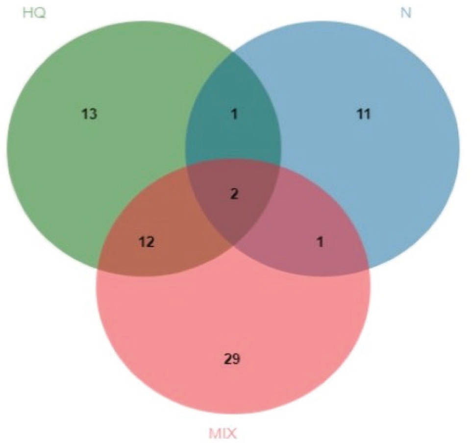
Figure 2
Venn diagrams comparing differentially expressed proteins obtained after treatment with nitrofurantoin (N), hydroquinone (HQ), and their mixture (MIX) after 8 h
Table 6
Complete Gene Ontology (GO) classification of differentially expressed proteins (DEPs) in the cells after treatment with the mixture of nitrofurantoin and the hydroquinone by biological processes
| GO biological process | No. of proteins in reference list* | Genes encoding differentially expressed proteins | P-value | False discovery rate |
|---|---|---|---|---|
| Glutamine catabolic process (GO:0006543) | 3 | GLS | 1.22×10⁻⁵ | 3.07×10⁻² |
| Glutamate biosynthetic process (GO:0006537) | 4 | GLS | 2.43×10⁻⁵ | 4.60×10⁻² |
| Cellular biosynthetic process (GO:0044249) | 3530 | CALR, UGGT1, GLS, ALDH18A1, INTS1, U2AF2, HSP90B1, TXNDC5, PDIA4, PDIA3, STOML2, HSD17B12, CCT5, PSIP1, RPLP0, MBOAT7, HACD2, HNRNPR | 2.13×10⁻⁵ | 9.75×10⁻³ |
| Glutamine family amino acid biosynthetic process (GO:0009084) | 15 | GLS, ALDH18A1 | 3.54×10⁻⁶ | 1.78×10⁻² |
| Protein folding in endoplasmic reticulum (GO:0034975) | 11 | CALR, HSP90B1, PDIA3 | 1.29×10⁻⁶ | 4.72×10⁻³ |
| Protein folding (GO:0006457) | 223 | CALR, UGGT1, HSP90B1, TXNDC5, PDIA4, PDIA3, CCT5 | 3.12×10⁻⁷ | 1.87×10⁻² |
| Protein maturation (GO:0051604) | 494 | CALR, UGGT1, HSP90B1, TXNDC5, PDIA4, PDIA3, STOML2, CCT6 | 6.00×10⁻⁶ | 2.27×10⁻² |
| Response to endoplasmic reticulum stress (GO:0034976) | 226 | CALR, UGGT1, HSP90B1, PDIA4, PDIA3, PTPN1 | 6.19× 10⁻⁶ | 4.59×10⁻² |