
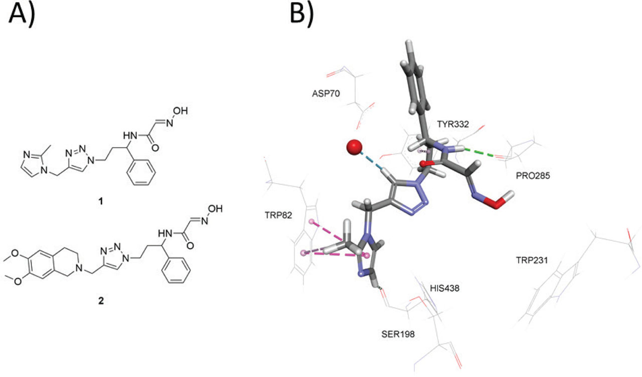
Figure 1
A) N-substituted 2-hydroxyiminoacetamides 1 and 2; B) crystallographic structure of oxime (R)-1 bound to human BChE (PDB: 6T9P). Non-covalent interactions are shown: hydrogen bonds (green dashed lines) and π interactions (magenta dashed lines). 1 – 2-hydroxyimino-N-(3-(4-((2-methyl-1H-imidazol-1-yl)methyl)-1H-1,2,3-triazol-1-yl)-1-phenylpropyl)acetamide; 2 – N-(3-(4-((6,7-dimethoxy-3,4-dihydroisoquinolin-2(1H)-yl)methyl)-1H-1,2,3-triazol-1-yl)-1-phenylpropyl)-2-(hydroxyimino)acetamide

Figure 2
Synthesis of the test compound N-(3-(4-((6,7-dimethoxy-3,4-dihydroisoquinolin-2(1H)-yl)methyl)-1H-1,2,3-triazol-1-yl)-1-phenylpropyl)-2-(hydroxyimino)acetamide. 2 – N-(3-(4-((6,7-dimethoxy-3,4-dihydroisoquinolin-2(1H)-yl)methyl)-1H-1,2,3-triazol-1-yl)-1-phenylpropyl)-2-(hydroxyimino) acetamide; 3 – N-(3-azido-1-phenylpropyl)-2-hydroxyiminoacetamide; 4 – 6,7-dimethoxy-2-(prop-2-yn-1-yl)-1,2,3,4-tetrahydroisoquinoline
Table 1
Reversible inhibition of human red blood cell acetylcholinesterase (AChE) and human plasma butyrylcholinesterase (BChE) by the test compound N-(3-(4-((6,7-dimethoxy-3,4-dihydroisoquinolin-2(1H)-yl)methyl)-1H-1,2,3-triazol-1-yl)-1-phenylpropyl)-2-(hydroxyimino)acetamide
| Enzyme | Acetylthiocholine (mmol/L) | Concentration range (µmol/L) | KI (µmol/L) |
|---|---|---|---|
| AChE | 0.1–1.0 | 25–75 | 49±4 |
| BChE | 0.1–1.0 | 5–30 | 18±1 |
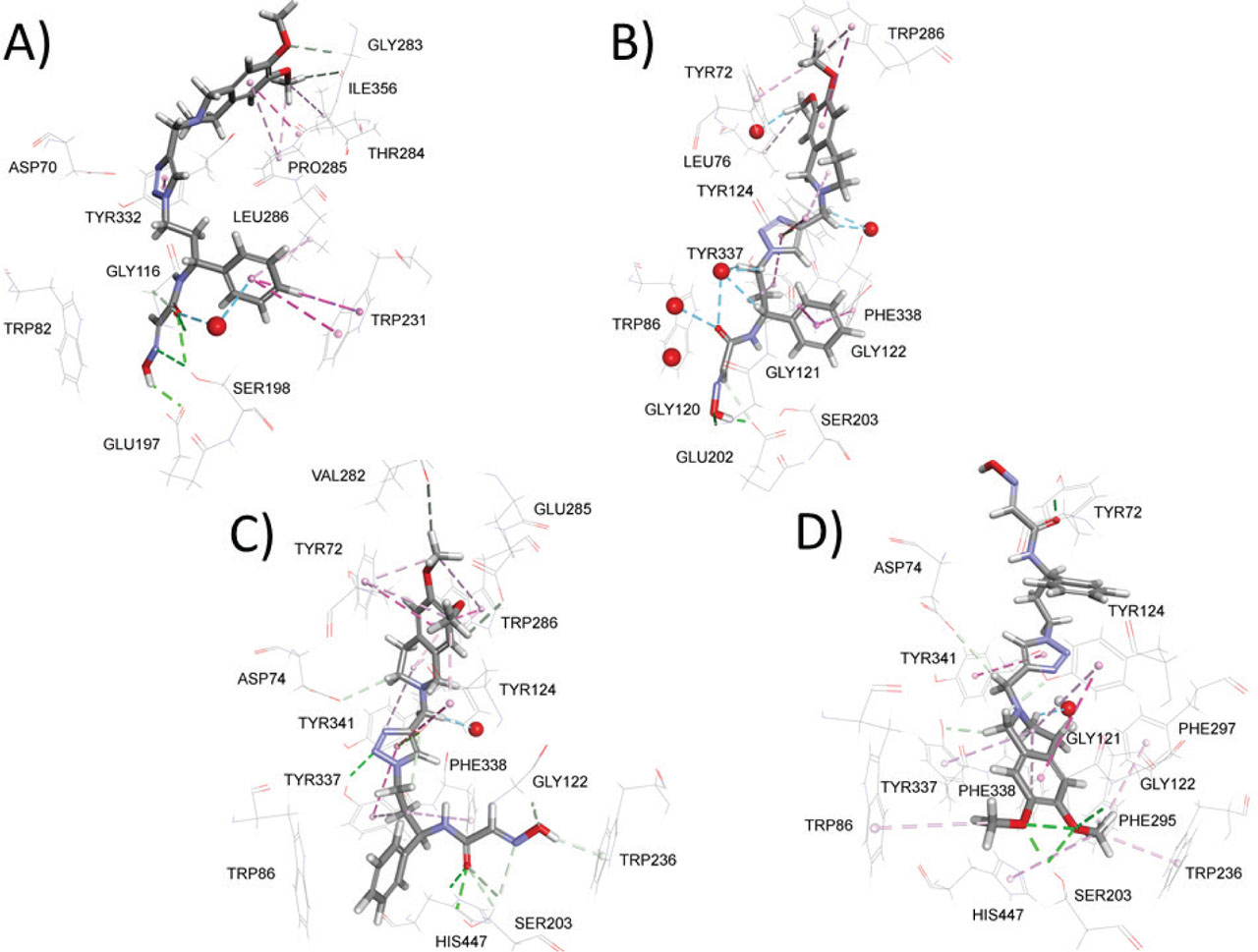
Figure 3
Model complexes of the test compound N-(3-(4-((6,7-dimethoxy-3,4-dihydroisoquinolin-2(1H)-yl)methyl)-1H-1,2,3-triazol-1-yl)-1-phenylpropyl)-2-(hydroxyimino)acetamide and human BChE (A) or human AChE (B–D). Dashed lines represent different types of non-bonding interactions (magenta – π-alkyl, π-π interaction; green – conventional hydrogen bond; light green – carbon hydrogen bond; blue – water hydrogen bond). Red spheres show only those water molecules (hydrogens hidden) predicted to interact with ligands