
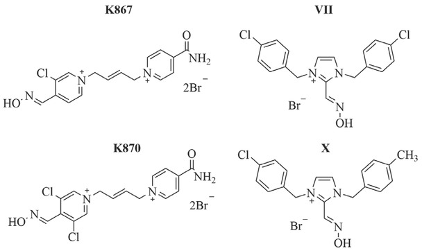
Figure 1
Structures of oximes tested in this study
Table 1
Primary antibodies (Cell Signaling Technology Inc, USA) used for Western blotting
| Antibody | Cat. No. | Host | Type | Dilution |
|---|---|---|---|---|
| Anti p-ERK (Thr202/Tyr204) | #4370 | Rabbit | Monoclonal | 1:2000 |
| Anti pNFκB p65 (Ser536) | #3033 | Rabbit | Monoclonal | 1:2000 |
| Anti p-p38 MAPK (Thr180/Tyr182) | #4511 | Rabbit | Monoclonal | 1:1000 |
| Anti p-STAT3 (Tyr705) | #9145 | Rabbit | Monoclonal | 1:1000 |
| Anti p-ACC (Ser79) | #3661 | Rabbit | Polyclonal | 1:1000 |
| Anti p-AMPKα (Thr172) | #2535 | Rabbit | Monoclonal | 1:1000 |
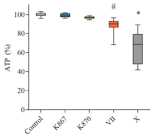
Figure 2
ATP in SH-SY5Y cells after 4 h exposure to selected oximes expressed as the percentage of control (untreated cells). Oxime concentrations: K867 (400 μM), K870 (15 μM), VII (250 μM) and X (200 μM). #p≤0.01; *p≤0.0001
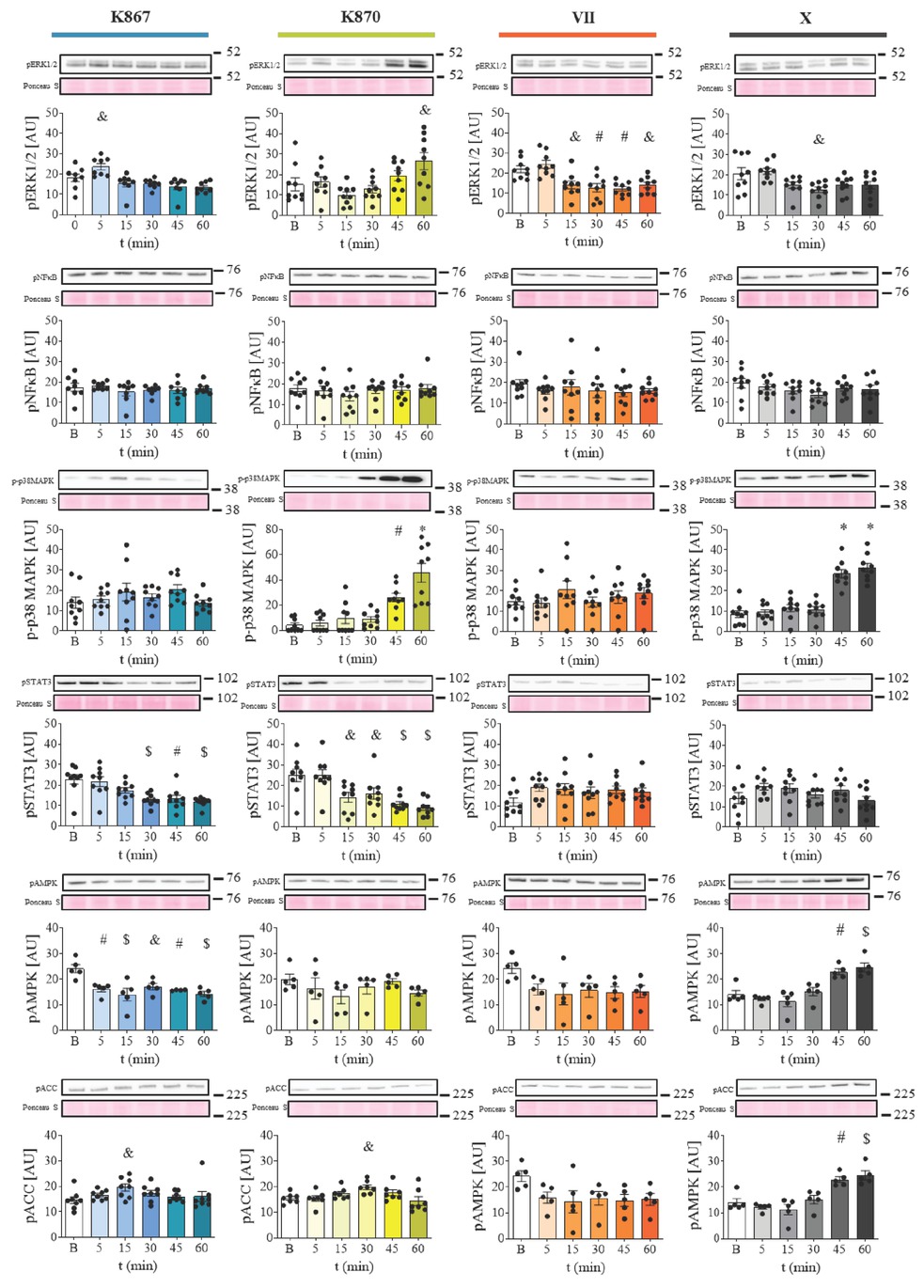
Figure 3
Time-dependent effects of the selected oximes K867 (400 μmol/L), K870 (15 μmol/L), VII (250 μmol/L), and X (200 μmol/L) on intracellular signalling in serum-starved SH-SY5Y cells over 5–60 min. Position towards bends and molecular weight markers in kDa are indicated on the right side of the blots and Ponceau S. Results are presented as means ± SD (n=9) &p≤0.05; #p≤0.01; $p≤0.001; *p≤0.0001. B– baseline; pERK1/2 – phospho-ERK1/2 (Thr202/Tyr204); pNFkB – phospho-NFκbeta p65 (Ser536); p-p38MAPK – phospho-p38MAPK (Thr180/Tyr182); p-STAT3 – phospho-STAT3 (Tyr705); p-ACC – phospho-ACC (Ser79); p-AMPKα – phospho-AMPKalpha (Thr172); t– time
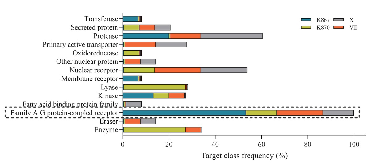
Figure 4
Target class frequencies of oximes K867, K870, VII, and X predicted by the SwissTargetPrediction engine. The dashed frame highlights the family of G-protein-coupled receptors as the most probable target of interaction
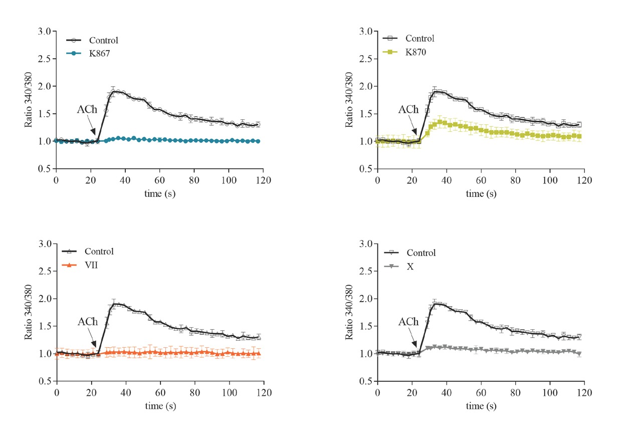
Figure 5
iCa2+ levels in SH-SY5Y cells before and after the addition of ACh (5 μmol/L) in the presence of oximes K867 (400 μmol/L), K870 (25 μmol/L), VII (250 μmol/L), and X (200 μmol/L) compared to control (not treated with the oximes)