
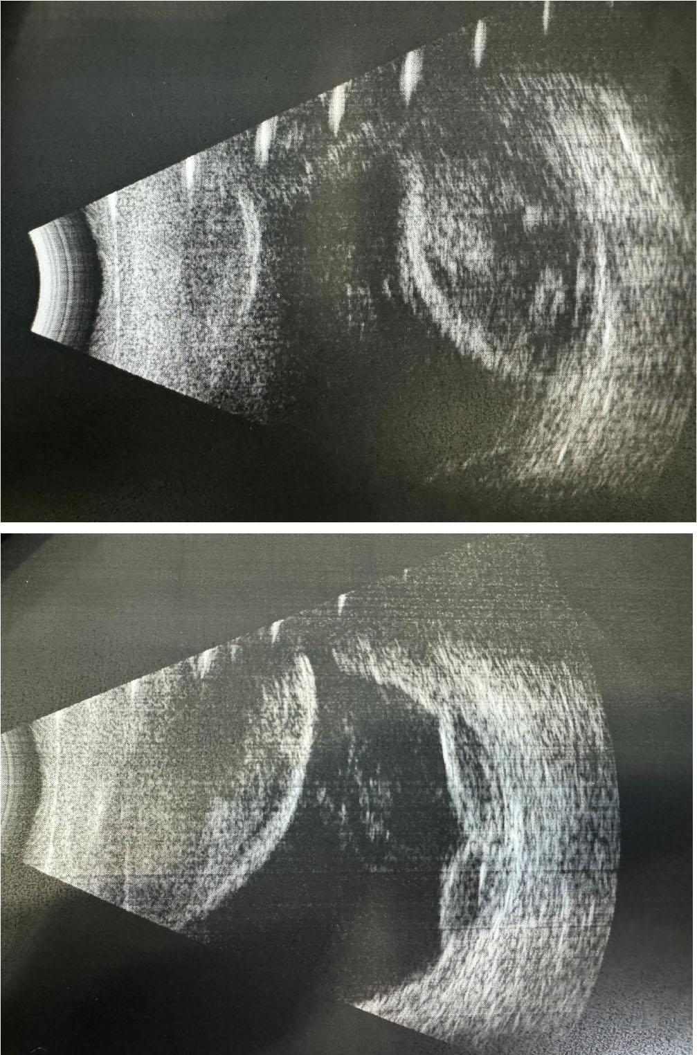
Figure 1.
B-projection ultrasound image of the OS, massive formations of the separated choroid, the so-called “kissing choroidal sign”
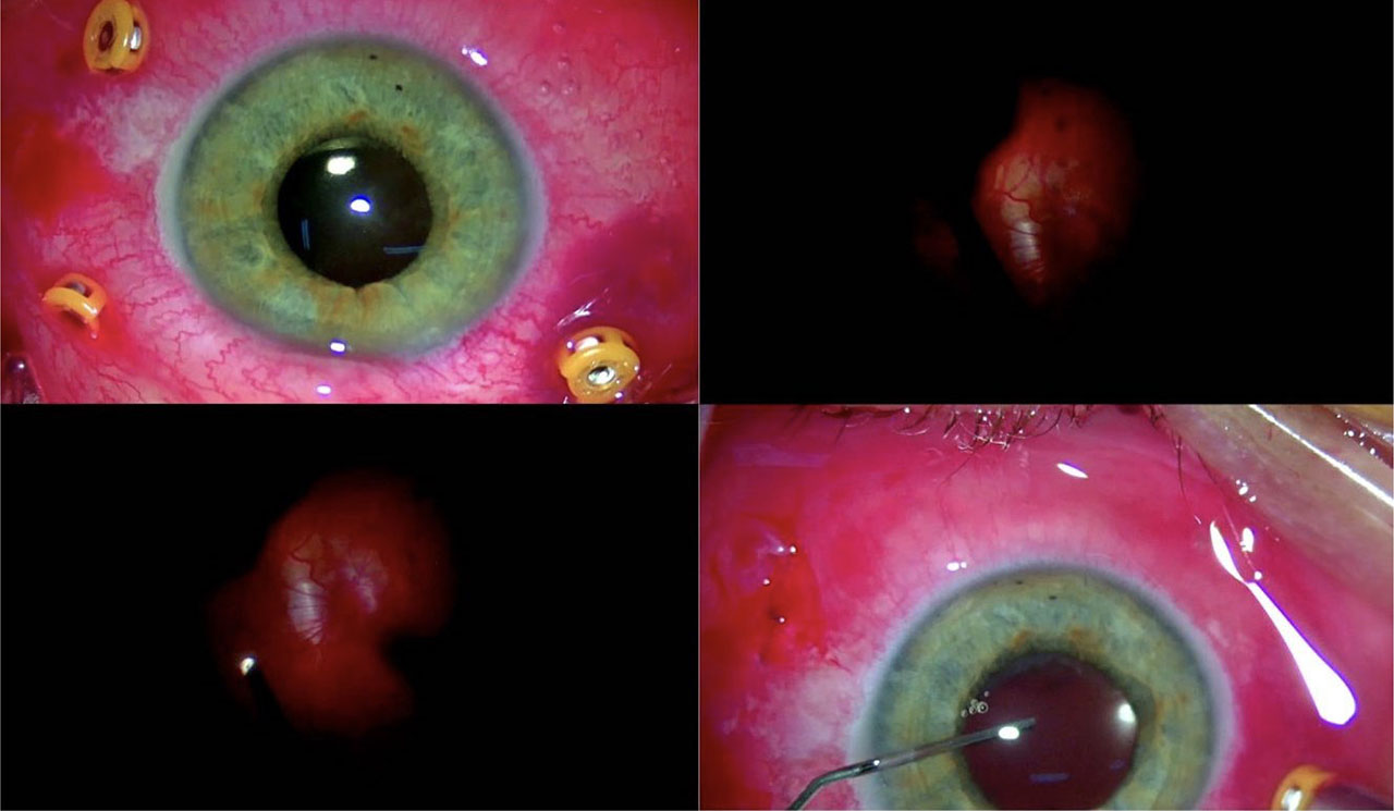
Figure 2.
PPV (23G) OS with mAndo iridotomy, partial evacuation of subretinal and subretinal blood, retinal endolaser, PFCL removal and silicone oil endotamponade
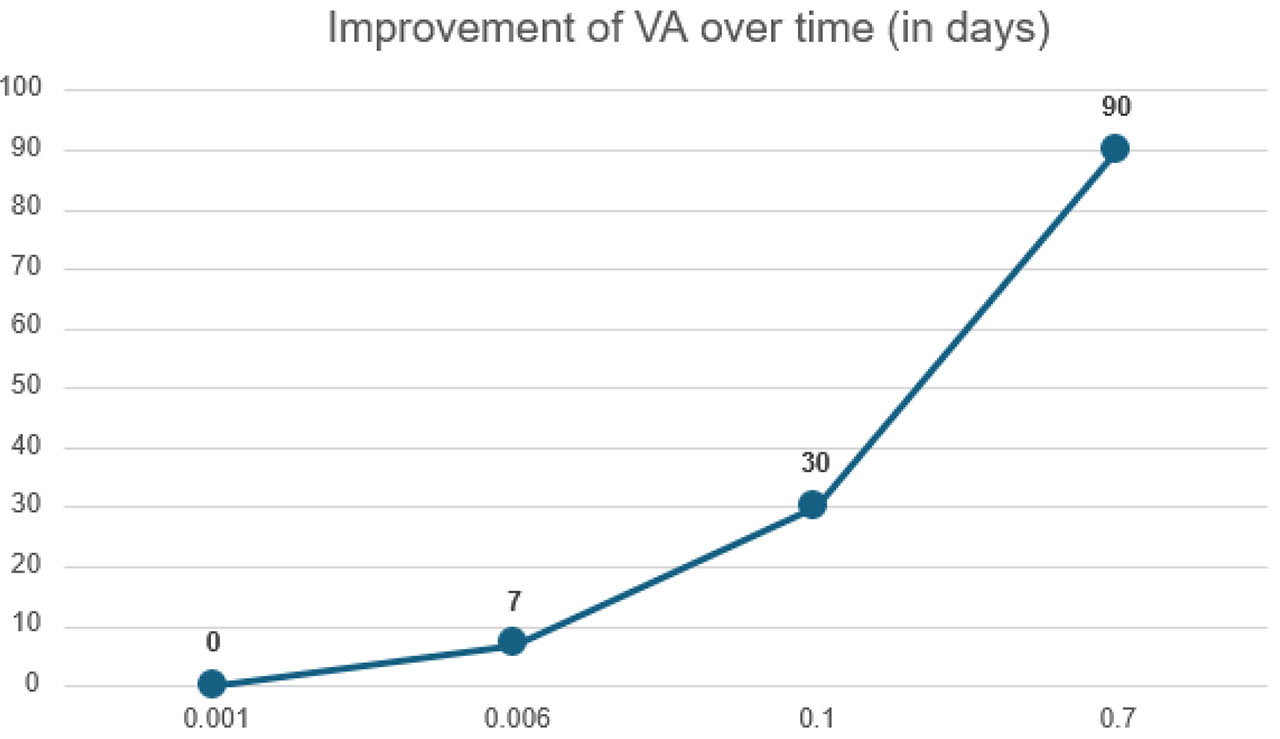
Figure 3.
Line graph of visual acuity versus time since surgery
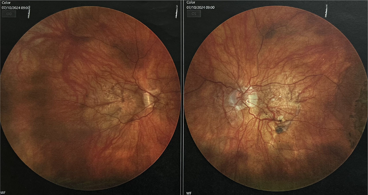
Figure 4.
Color photograph of the fundus of the OD and SL; in the OS, the endotamponade with silicone oil is visible, as well as the applied retina in all quadrants
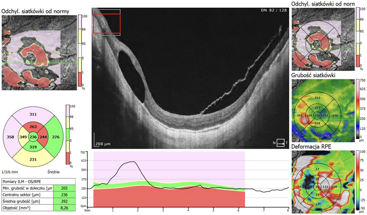
Figure 5.
Optical coherence tomography study of the OS 5 months after the PPV procedure