
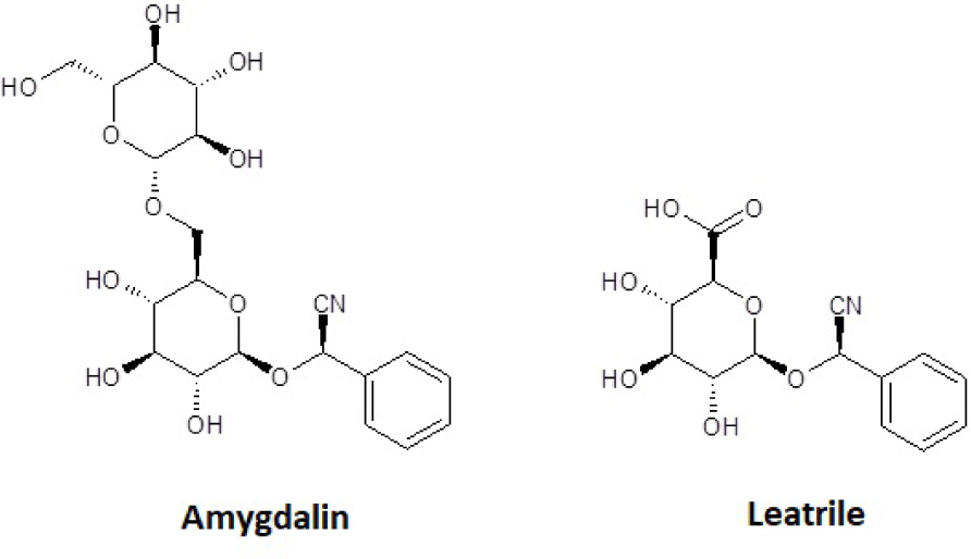
Fig. 1
Comparison of the chemical structure of amygdalin and laetrile.
Table 1
Human cell lines used in the study.
| Cells characteristics | Cel lines* | |||
|---|---|---|---|---|
| T-47D | MCF-7 | MDA-MB-231 | HFF-1 | |
| Morphology | Ephithelial | Ephithelial | Ephithelial | Fibroblasts |
| Tissue | Mammary gland/derived from metastatic site: pleural effusion | Mammary gland/derived from metastatic site: pleural effusion | Mammary gland/derived from metastatic site: pleural effusion | Skin: foreskin |
| Disease | Ductal carcinoma/estrogen-dependent | Adenocarcinoma/estrogen-dependent | Adenocarcinoma/estrogen-independent | Normal |
| Donor | 54 years adult/female | 69 years adult/female | 51 years adult/female | Newborn/male |
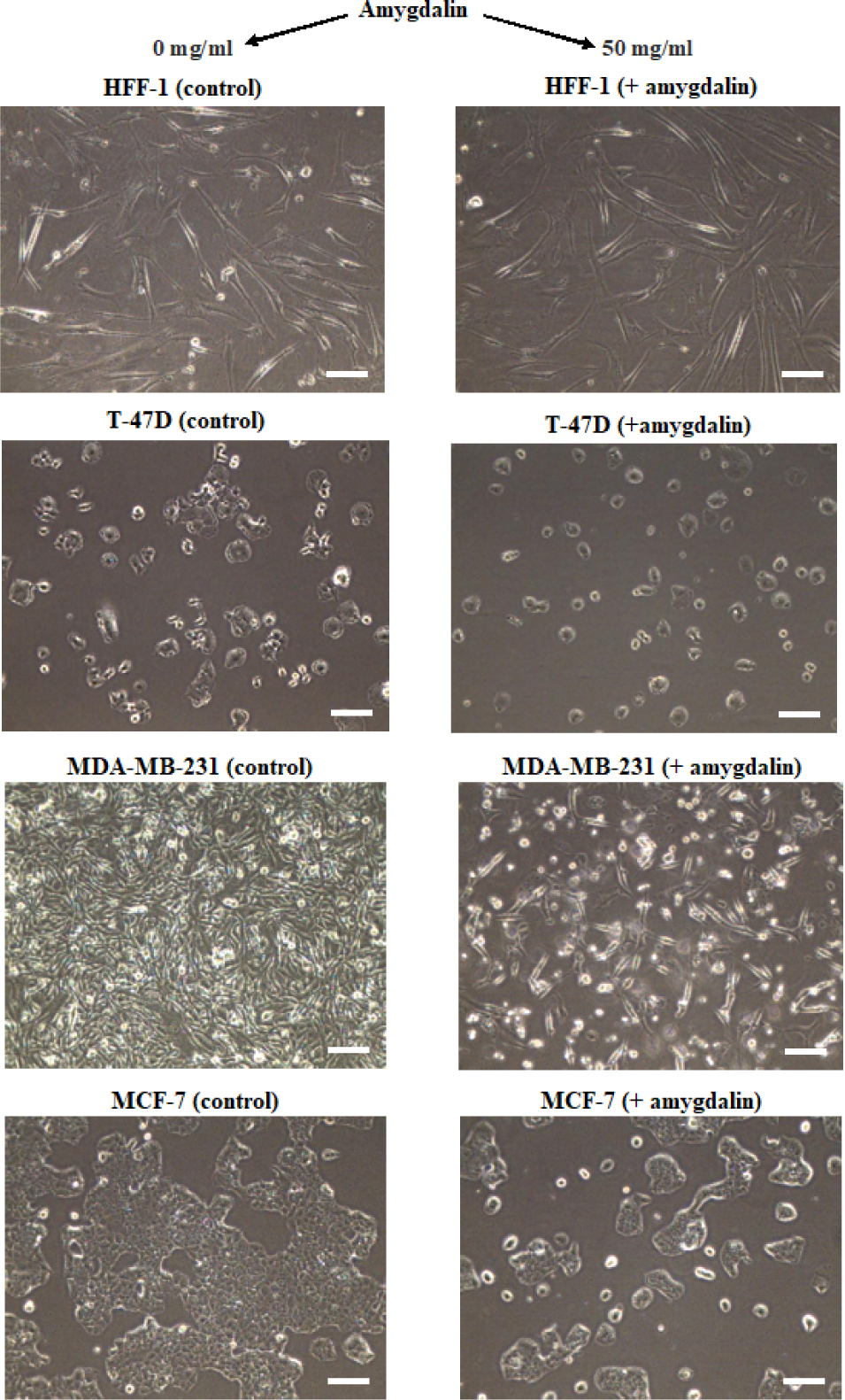
Fig. 2
Comparison of the confluence degree between the cell cultures treated with amygdalin at a concentration of 50 mg/ml and the control cultures – not treated with amygdalin. The microscopic image [100x] was collected after 48 hours of cell cultures incubation. Scale bar = 100 μm.
Table 2
Nucleotide sequences of the primers used in the RT-qPCR reaction.
| Primer name | Nucleotide sequence | Transcript detected | GenBank database access number |
|---|---|---|---|
| KAS3F | 5′-GGCCTGCCGTGGTACAGAACTGG-3′ | CASP3 mRNA | NM032991 |
| KAS3R | 5′-AGCGACTGGATGAACCAGGAGCCA-3′ | ||
| BAXF | 5′-CCTGTGCACCAAGGTGCCGGAACT-3′ | BAX mRNA | L22474 |
| BAXR | 5′-CCACCCTGGTCTTGGATCCAGCCC-3′ | ||
| BCL-2F | 5′-TTGTGGCCTTCTTTGAGTTCGGTG-3′ | BCL2 mRNA | M14745 |
| BCL-2R | 5′-GGTGCCGGTTCAGGTACATCAGTCA-3′ | ||
| 18SrRNAF | 5′-CAGTTATGGTTCCTTTGGTCGCTC-3′ | 18S rRNA | M10098 |
| 18SrRNAR | 5′-GTTGATAGGGCAGACGTTCGAATG-3′ |
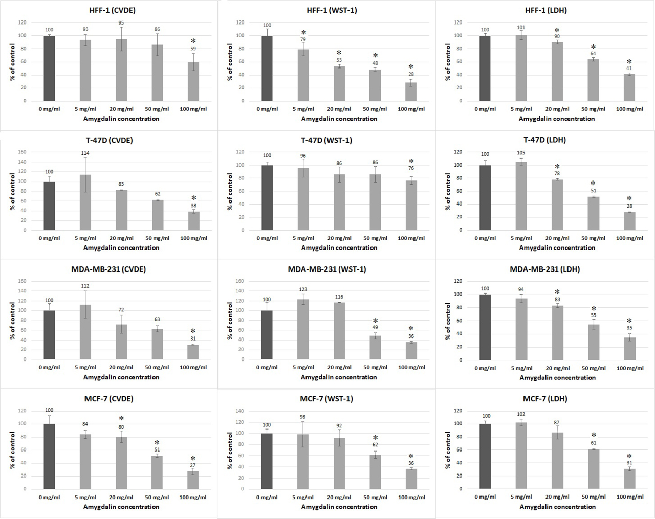
Fig. 3
The effect of different doses of amygdalin (0 – 100 mg/ml) on the percentage of attached, viable cells (CVDE test), on the metabolic activity of cells (WST-1 test) and on the percentage of dying cells (LDH test) in the HFF-1, T-47D, MDA-MB-231, and MCF-7 in vitro cultures. Statistical significance means p <0.05 (*).
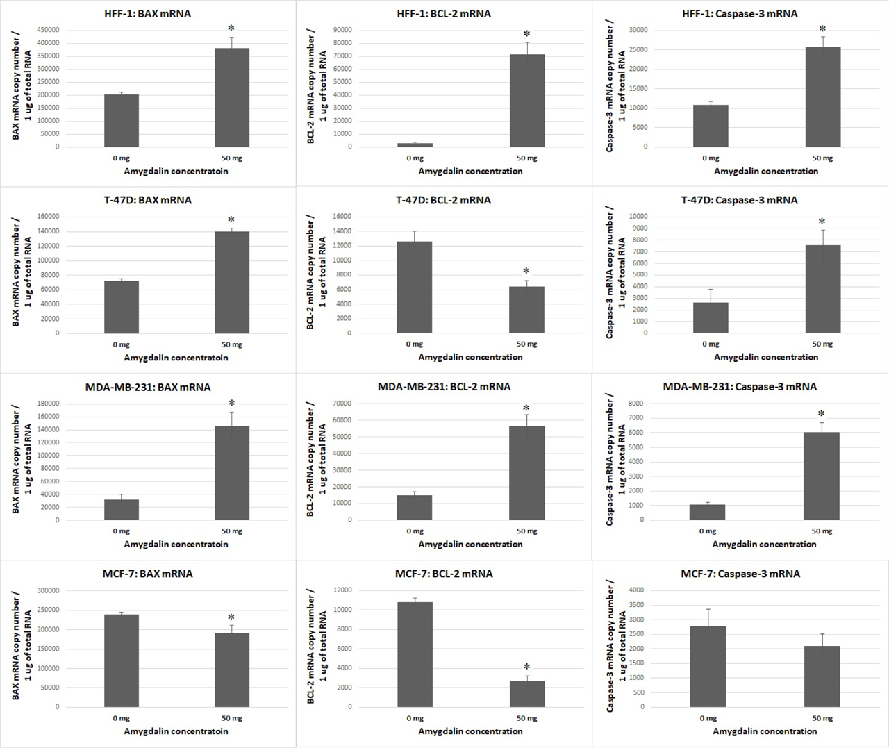
Fig. 4
The effect of amygdalin at a concentration of 50 mg/ml on the BAX, BCL2, and caspase-3 mRNA level in human breast cancer cells (T-47D, MDA-MB-231, MCF-7) and normal dermal fibroblasts in vitro compared to control (cell cultures not treated with amygdalin). Statistical significance means p <0.05 (*).
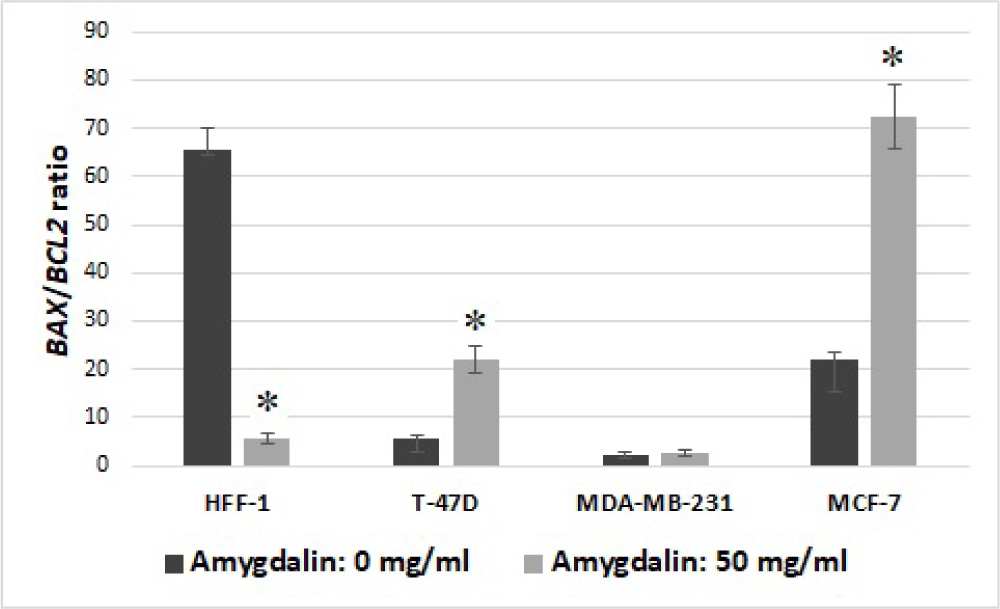
Fig. 5
The BAX/BCL2 ratio (BAX mRNA copy number/BCL-2 mRNA copy number) in culture cells treated with amygdalin (at concentration of 50 mf/ml) compared to control (culture cells not treated with amygdalin). Statistical significance means p <0.05 (*).