
FIGURE 1
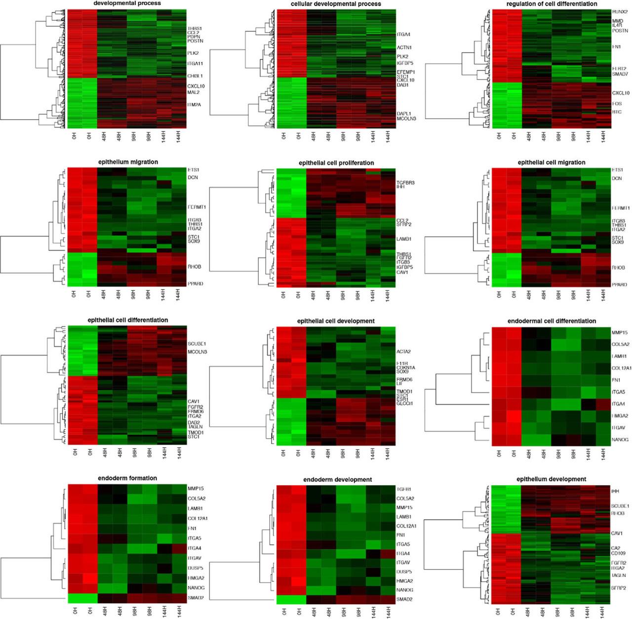
FIGURE 2
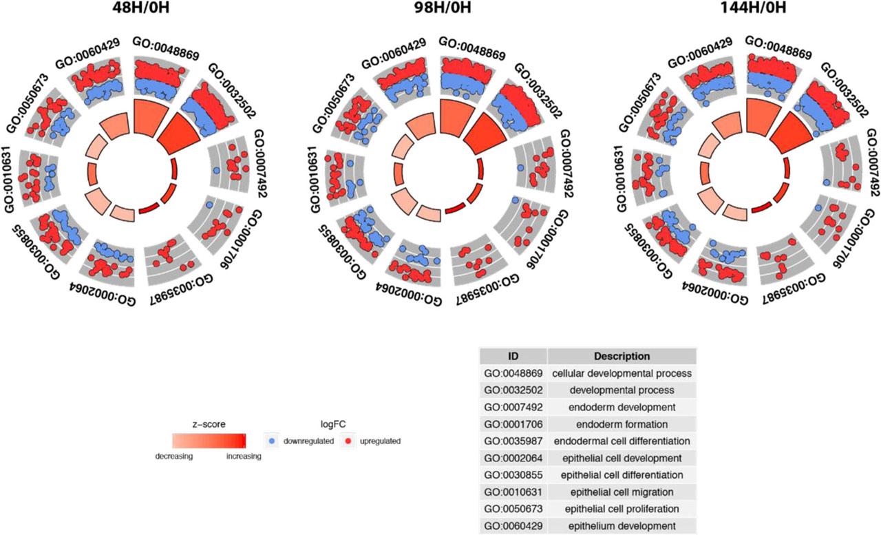
FIGURE 3
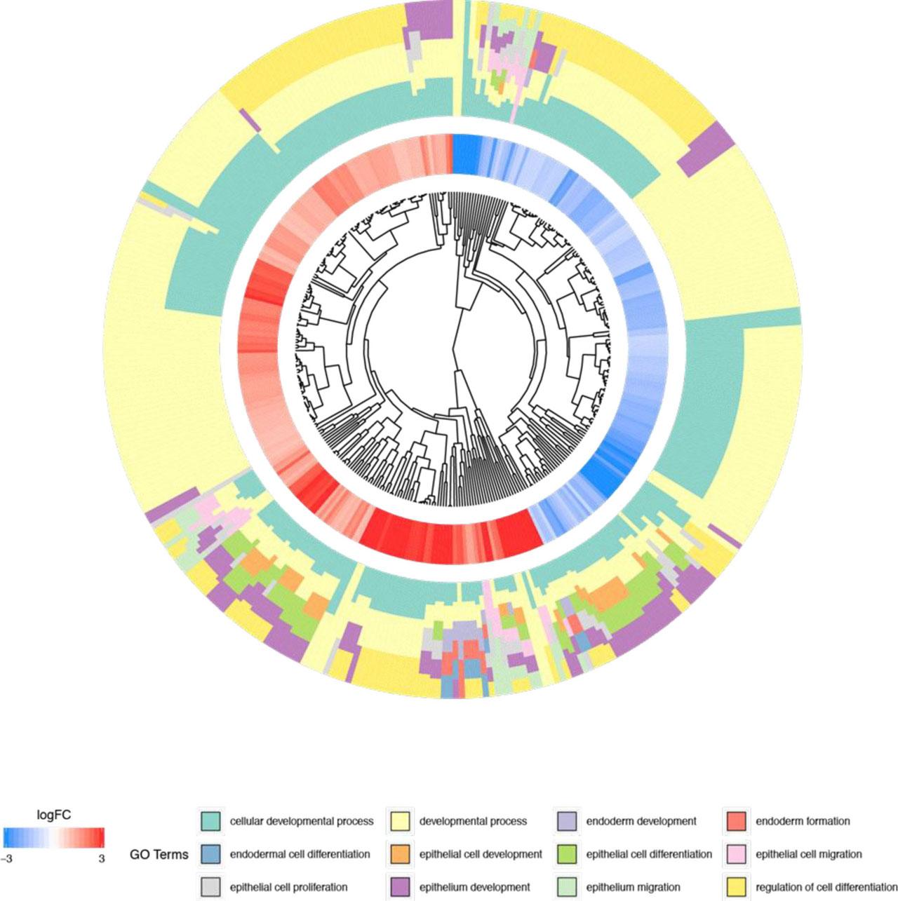
FIGURE 4
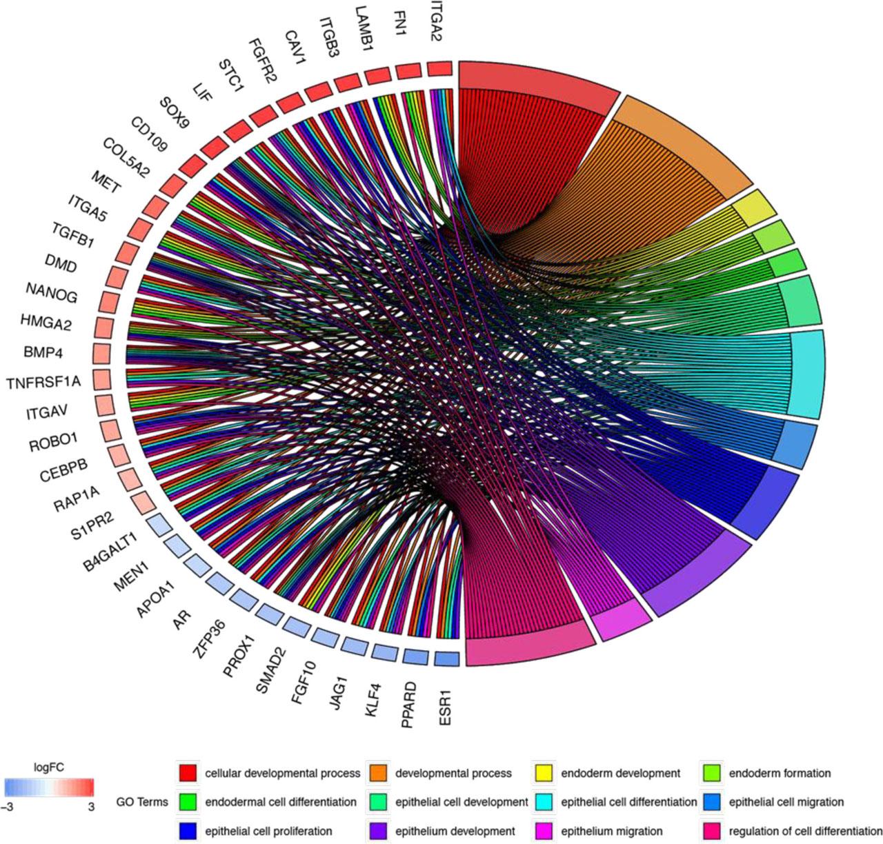
FIGURE 5
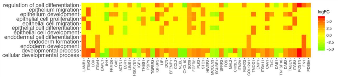
FIGURE 6
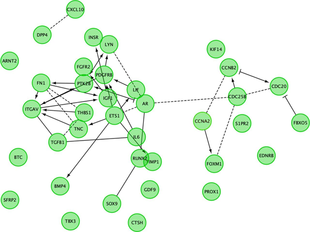
FIGURE 7
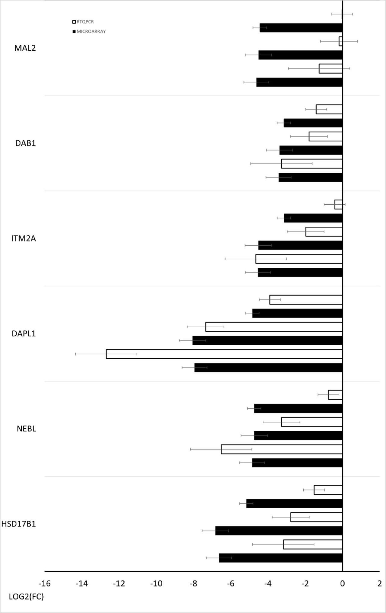
The 6 most downregulated genes involved in regulation of granulosa cells differentiation towards endodermal and epithelial tissues were validated using RT-qPCR, 168h/0h
| GENE NAME | GENE SYMBOL | FOLD CHANGE 48H/0H | ADJ. P. VAL. |
|---|---|---|---|
| integral membrane protein 2A | ITM2A | -8.85 | <0.05 |
| disabled homolog 1 (Drosophila) | DAB1 | -8.91 | <0.05 |
| mal, T-cell differentiation protein 2 | MAL2 | -21.75 | <0.05 |
| nebulette | NEBL | -26.72 | <0.05 |
| death associated protein-like 1 | DAPL1 | -28.69 | <0.05 |
| hydroxysteroid (17-beta) dehydrogenase 1 | HSD17B1 | -35.75 | <0.05 |
Primer sequences (5′-3′)
| GENE NAME | GENE SYMBOL | PRIMER SEQUENCES (5′-3′) |
|---|---|---|
| integral membrane protein 2A | ITM2A | TCTCGTAGGCCTTTCCTTCA |
| disabled homolog 1 (Drosophila) | DAB1 | TACGTTTGTGGGAAGGAAGG |
| mal, T-cell differentiation protein 2 | MAL2 | AGGATGGGTCATGTTCGTGT |
| nebulette | NEBL | CAAACCCTTCAAGGCTACCA |
| death associated protein-like 1 | DAPL1 | CCTGCTCTGGAGAAGGTCAC |
| hydroxysteroid (17-beta) dehydrogenase 1 | HSD17B1 | GTGTCAGAGGCTTGCTAGGG |