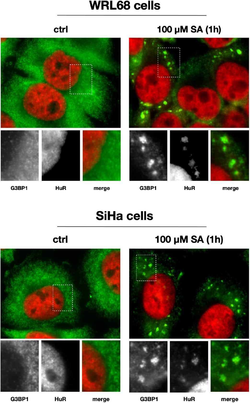
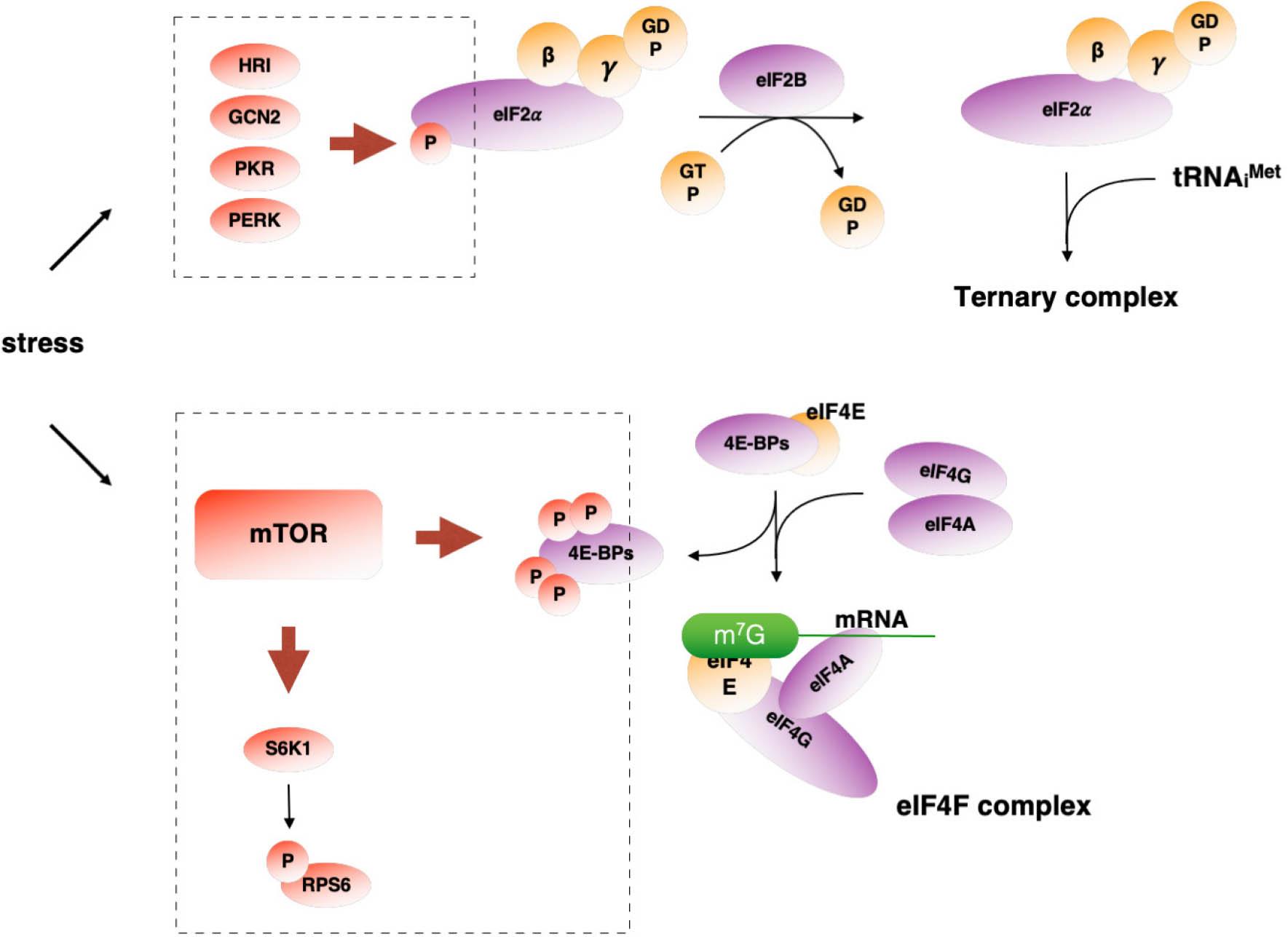
Have a personal or library account? Click to login
Molecular structure of stress granules and their role in the eukaryotic cell Cover

Molecular structure of stress granules and their role in the eukaryotic cell

Open Access
|Mar 2021

Figures & Tables

Figure 1

An example of stress granules formation in two types of cells (WRL68 and SiHa) using immunofluorescence method. This figure shows two stress granules markers: G3BP1 and HuR as well as nucleus in cells under normal growth conditions (ctrl) and the formation of stress granules under stressful conditions caused by sodium arsenite (SA)

Figure 2

Two mechanisms for regulating protein biosynthesis. The top diagram shows the mechanism based on the phosphorylation of the eIF2α factor, which is involved in the correct binding of the initiator tRNA to the ribosome. When one of the kinases is activated, the eIF2α factor is phosphorylated, which leads to blocking the formation of the triple complex and stopping the initiation process. The second mechanism is related to the mTOR signaling pathway, which regulates the phosphorylation of 4E-BP and RPS6 (ribosomal protein S6) proteins. Under normal growth conditions, mTOR phosphorylates 4E-BP proteins, preventing them from binding to eIF4E, whereby the latter may form the active eIF4F initiation complex. Inhibition of mTOR activity causes hypophosphorylation of the 4E-BP proteins which bind to eIF4E in this state, thereby blocking the formation of the eIF4F complex
Language: English
Page range: 33 - 41
Submitted on: Feb 12, 2021
Accepted on: Mar 11, 2021
Published on: Mar 30, 2021
Published by: Foundation for Cell Biology and Molecular Biology
In partnership with: Paradigm Publishing Services
Publication frequency: 4 issues per year

© 2021 Paulina Pietras, Marta Leśniczak, Mateusz Sowiński, Witold Szaflarski, published by Foundation for Cell Biology and Molecular Biology
This work is licensed under the Creative Commons Attribution-NonCommercial-NoDerivatives 4.0 License.