
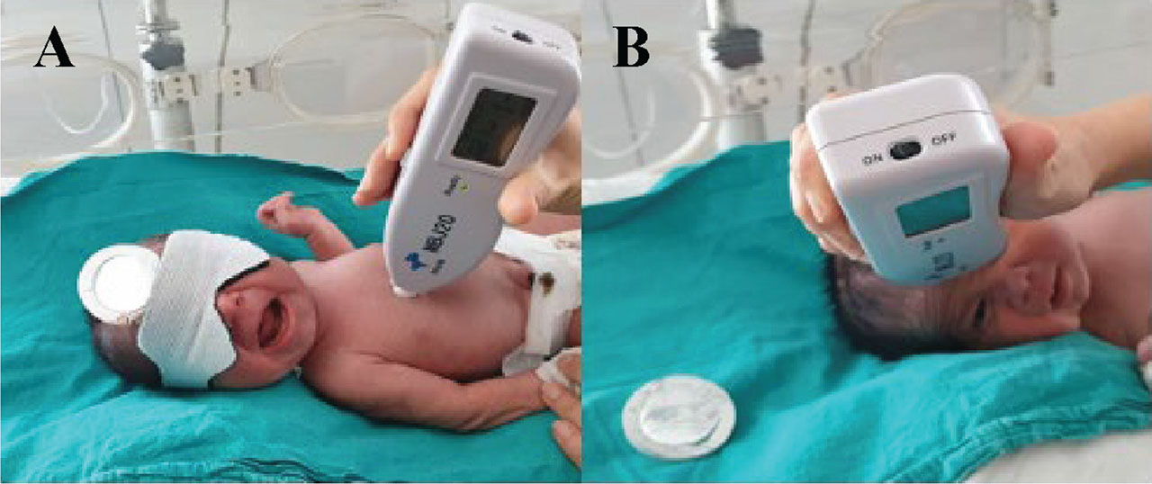
Figure 1.
TcB measurements after PT. (A) TcB measurement at the exposed area and (B) TcB measurement under the covered shield. The neonate’s face is shown with full, explicit written informed consent obtained from the parents for publication of this identifiable image. The parents were informed about the nature, purpose, and extent of the publication, including digital distribution. PT, phototherapy; TcB, transcutaneous bilirubin.
Table 1.
Epidemiological characteristics of the neonates receiving PT for hyperbilirubinemia
| Characteristics | N (%) | |
|---|---|---|
| Gestational age (weeks), median (range) | 38 (31–42) | |
| Birth weight (g), mean ± SD | 3020 ± 556 | |
| Sex | ||
| Female | 36 (51.4) | |
| Male | 34 (48.6) | |
| Maternal age (years), mean ± SD | 26.3 ± 5.7 | |
| Maternal disease status | ||
| Yes | 8 (11.4) | |
| No | 60 (88.6) | |
| Mode of delivery | ||
| NSVD | 51 (72.9) | |
| C/S | 19 (27.1) | |
| Age at admission (days), median (range) | 2 (1–13) | |
| Length of stay (days), median (range) | 3 (2–27) | |
| Blood incompatibility | ||
| None | 25 (35.8) | |
| ABO incompatibility | 41 (58.6) | |
| Rh incompatibility | 3 (4.2) | |
| ABO + Rh incompatibility | 1 (1.4) | |
| DC test | ||
| Negative | 47 (67.2) | |
| Positive | 23 (32.8) | |
| Pathological weight loss | ||
| Yes | 10 (14.3) | |
| No | 60 (85.7) | |
| Cephalic hematoma | ||
| Yes | 2 (2.9) | |
| No | 68 (97.1) |
Table 2.
Comparison of TSB and TcB levels from covered and exposed skin measured before, during, and after PT
| Before PT | At 24th hour of PT | 8 h after discontinuation of PT | |
|---|---|---|---|
| TSB | 11.91 ± 4.48 | 7.67 ± 2.34 | 7.90 ± 2.09 |
| Covered skin TcB | 11.11 ± 3.01 | 7.07 ± 2.25 | 8.61 ± 2.39 |
| Exposed skin TcB | 11.14 ± 3.11 | 4.13 ± 1.88 | 6.85 ± 2.44 |
Table 3.
Correlation between TSB values and TcB values measured from covered and exposed skin
| Before PT ICC (95% CI) | After 24 h of PT ICC (95% CI) | 8 h after PT ICC (95% CI) | |
|---|---|---|---|
| TSB vs. exposed skin TcB | 0.667 (0.513–0.780) | 0.229 (0.092–0.544) | 0.432 (0.200–0.613) |
| TSB vs. covered skin TcB | 0.665 (0.508–0.778) | 0.520 (0.326–0.672) | 0.537 (0.339–0.687) |
| Exposed vs. covered skin TcB | 0.828 (0.737–0.890) | <0.245 (0.095–0.542) | 0.557 (0.056–0.784) |
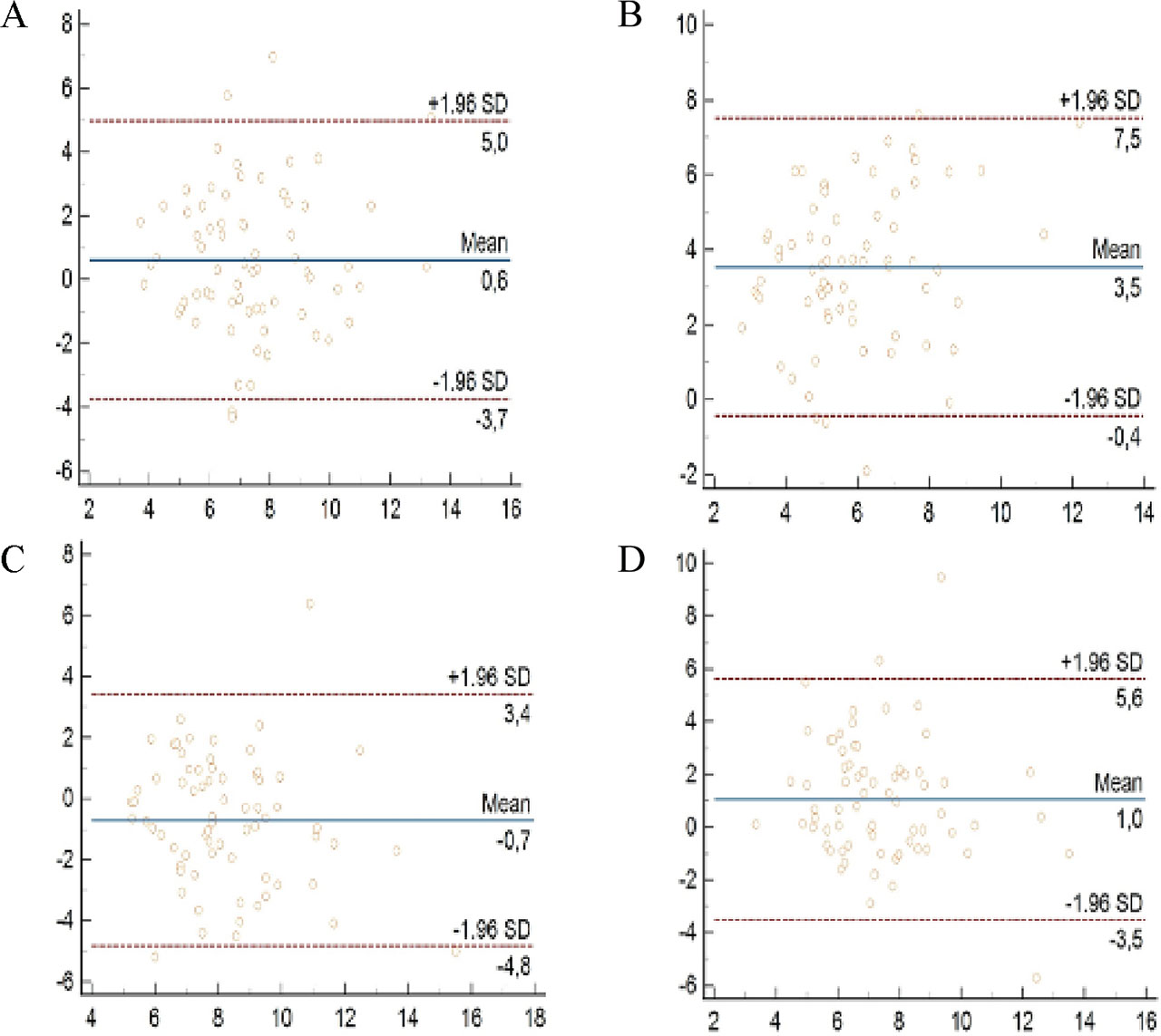
Figure 2.
Correlation between TSB and covered skin TcB (A) and exposed skin TcB (B) measurement after 24 h of PT. Correlation between TSB and covered skin TcB (C) and exposed skin TcB (D) measurements 8 h after discontinuing PT. The Bland-Altman plot shows the average of the two methods on the X-axis and the absolute difference between the two methods on the Y-axis. PT, phototherapy; TcB, transcutaneous bilirubin; TSB, total serum bilirubin.