
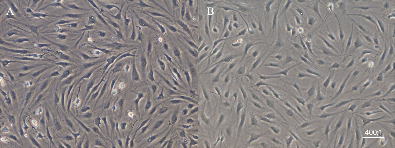
Figure 1
Morphology of chondrocytes in the 2 groups (×100). A. Chondrocytes in the experimental group were cultured with SDF-1 for 48 h. B. Morphology of chondrocytes in the control group. SDF-1, stromal cell-derived factor-1.
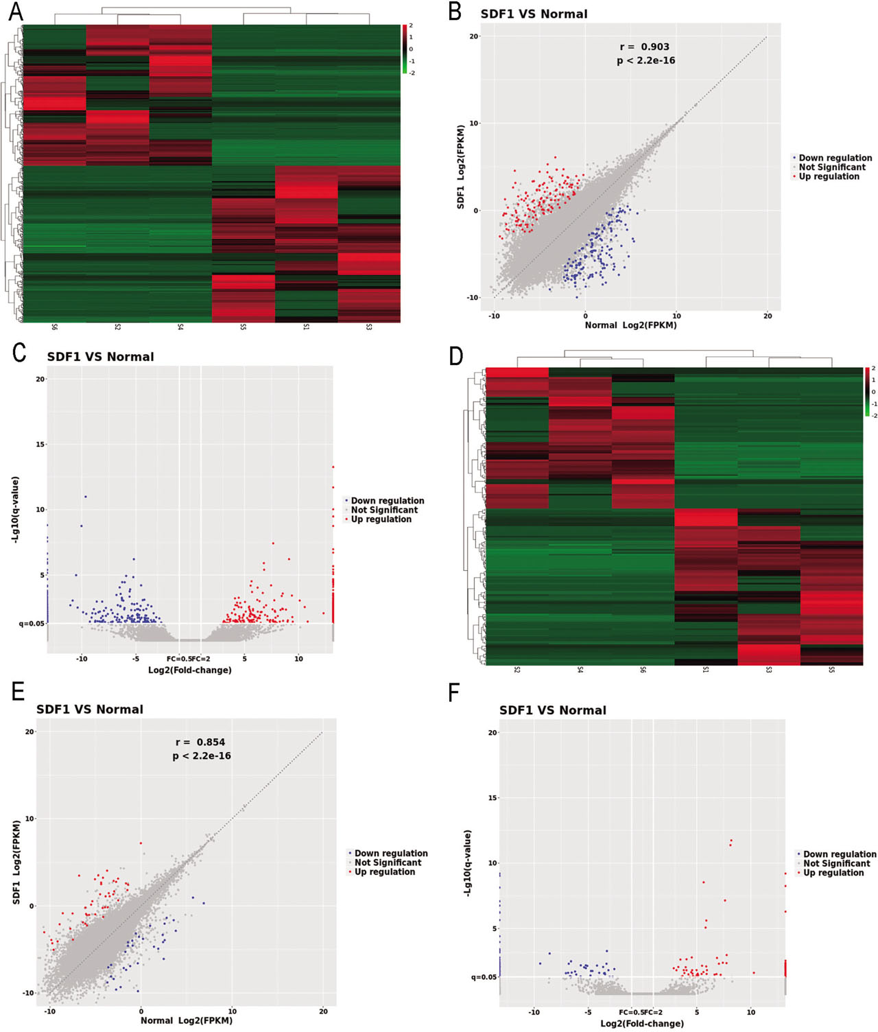
Figure 2
Analysis of miRNAs and lncRNAs. A. Heatmap of the differentially expressed (DE) miRNAs. B. Scatter plot of the DE miRNAs. C. Volcano plot of the DE miRNAs. D. Heatmap of the DE lncRNAs. E. Scatter plot of the DE lncRNAs. F. Volcano plot of the DE lncRNAs. In the heatmap, red represents upregulated miRNAs or lncRNAs, and green represents downregulated miRNAs or lncRNAs. In the scatter plot, the X and Y values are the average normalized signal values, shown on a log2 scale. The red and green lines were set as FC lines with a default change of 2.0. Red points (FC >2) indicate upregulated miRNAs or lncRNAs, and blue points (FC ≤2) indicate downregulated miRNAs or lncRNAs. In the volcano plot, the X-axis is the FC (log2), and the Y-axis is P (−log10). Red points (FC >2) indicate upregulated miRNAs or lncRNAs, and blue points (FC ≤2) indicate downregulated miRNAs or lncRNAs. DE, differentially expressed; FC, fold change; FPKM, fragments per kilobase of transcript per million mapped fragments; lncRNAs, long noncoding ribonucleic acids; miRNAs, microRNAs; SDF1, stromal cell-derived factor-1 treated articular chondrocytes. Normal indicates articular chondrocytes untreated with SDF-1.
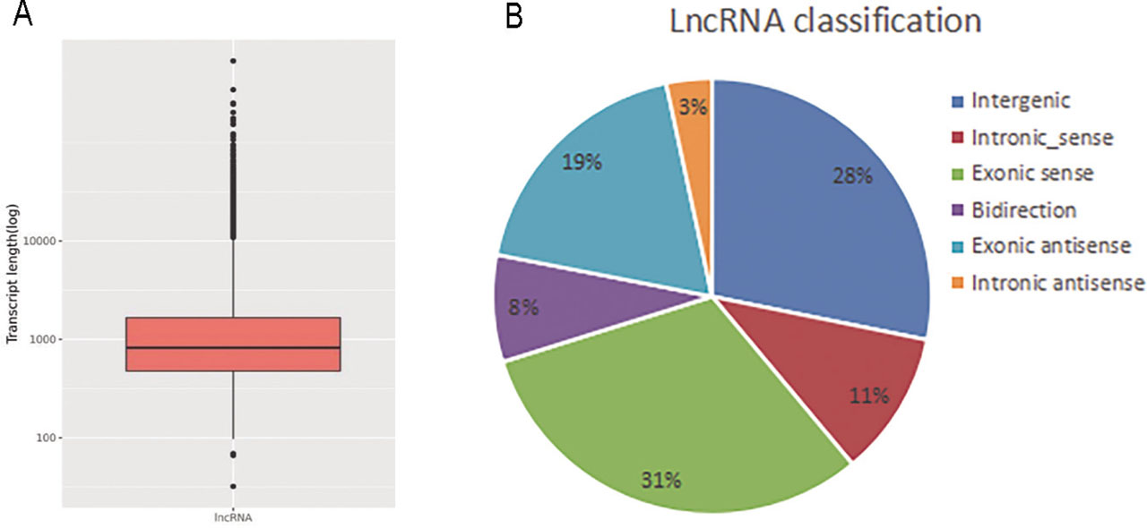
Figure 3
Expression signatures of dysregulated lncRNAs in SDF-1-induced articular chondrocyte degeneration. A. Length distribution showed that dysregulated lncRNAs were mainly concentrated between 700 bp and 3000 bp. B. Differential lncRNAs were classified according to their genomic architecture.
Table 1
Top 10 most upregulated and most downregulated lncRNAs in chondrocytes from the SDF-1-induced model of articular chondrocyte degeneration
| Upregulated lncRNAs | Downregulated lncRNAs | ||||
|---|---|---|---|---|---|
| lncRNA ID | P | FC | lncRNA ID | P | FC |
| NONHSAT094312.2 | 5.51E–05 | 10.27 | NONHSAT246243.1 | 4.01E–06 | 9.44 |
| NONHSAT060379.2 | 3.42E–17 | 8.19 | NONHSAT217441.1 | 3.97E–07 | 8.57 |
| NONHSAT207507.1 | 1.60E–16 | 8.12 | NONHSAT238505.1 | 6.22E–05 | 7.07 |
| NONHSAT166467.1 | 5.44E–07 | 7.77 | NONHSAT258030.1 | 5.57E–05 | 6.86 |
| NONHSAT198879.1 | 3.39E–06 | 7.75 | NONHSAT176410.1 | 4.59E–06 | 6.81 |
| NONHSAT248596.1 | 1.21E–11 | 7.61 | NONHSAT022132.2 | 0.000138 | 6.68 |
| NONHSAT152279.1 | 2.89E–06 | 7.41 | NONHSAT119402.2 | 2.11E–06 | 6.59 |
| NONHSAT000091.2 | 9.96E–05 | 7.21 | NONHSAT022138.2 | 0.000169 | 6.58 |
| ENST00000559458 | 4.66E–06 | 7.05 | NONHSAT229871.1 | 5.25E–05 | 6.53 |
| NONHSAT038052.2 | 1.00E–06 | 6.87 | NONHSAT225394.1 | 4.07E–05 | 6.23 |
[i] FC, fold change; SDF-1, stromal cell-derived factor-1.
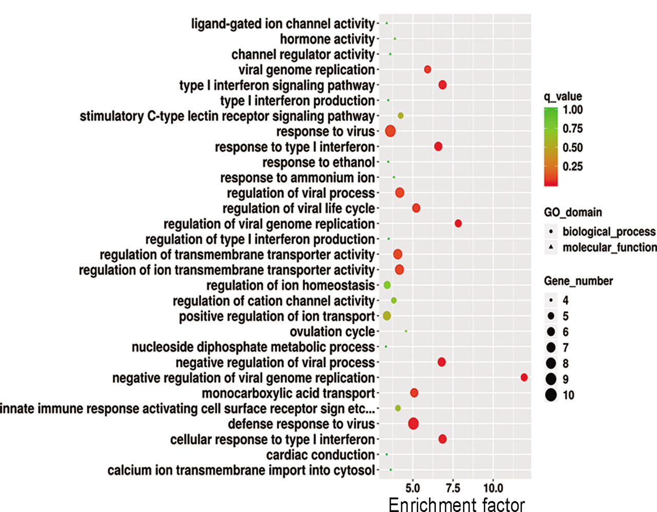
Figure 4
GO analysis of differentially expressed genes. GO annotations of mRNAs with top 30 enrichment scores. The circles represent biological processes; the triangles represent cell components; and the squares represent MF. GO, gene ontogeny; MF, molecular functions.
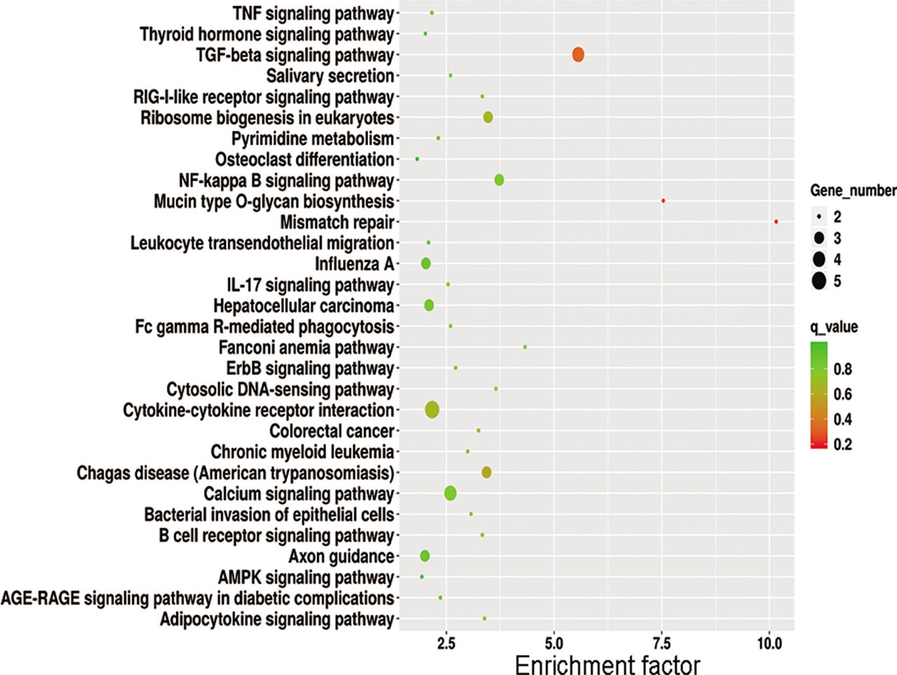
Figure 5
KEGG signaling pathway analysis of differentially expressed genes. Top 30 for KEGG enrichment. KEGG, Kyoto Encyclopedia of Genes and Genomes.
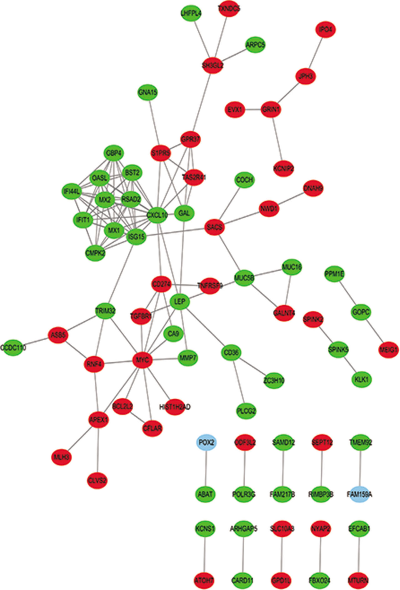
Figure 6
PPI network analysis of the top 10 differentially expressed genes. Nodes represent genes for the proteins indicated. Lines indicate interactions between genes. Red indicates upregulated genes, and green indicates downregulated genes. PPI, protein–protein interaction.
Table 2
PPI network core (top 10) genes in the SDF-1-induced model of articular chondrocyte degeneration
| Protein | Degree | Eccentricity | Edge count |
|---|---|---|---|
| CXCL10 | 16 | 4 | 16 |
| ISG15 | 12 | 4 | 12 |
| MYC | 11 | 5 | 11 |
| MX1 | 10 | 5 | 10 |
| OASL | 10 | 5 | 10 |
| IFIT1 | 10 | 5 | 10 |
| RSAD2 | 10 | 5 | 10 |
| MX2 | 10 | 5 | 10 |
| IFI44L | 10 | 5 | 10 |
| BST2 | 8 | 5 | 8 |
[i] BST2, bone marrow stromal tetherin antigen 2; CXCL10, interferon γ inducible CXC 10 kDa chemokine chemotactic for monocytes and T-lymphocytes; IFI44L, interferon-induced protein 44 like protein; IFIT1, interferon-induced with tetratricopeptide repeats 1 protein; ISG15, interferon-α-stimulated gene 15-kDa protein; MX1, interferon inducible myxovirus resistance protein MxA p78; MX2, second interferon-induced myxovirus resistance 2 protein MxB p78; MYC, v-myc myelocytomatosis viral oncogene homolog protein; OASL, p59 2′,5′-oligoadenylate synthetase-like protein; PPI, protein–protein interaction; RSAD2, radical S-adenosyl methionine domain containing 2 protein; SDF-1, stromal cell-derived factor-1.