
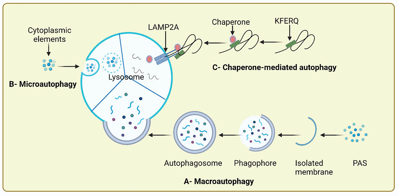
Figure 1
Schematic of autophagic routes. (A) Macroautophagy (commonly named autophagy): the autophagosome comprising several cytosolic polypeptides and proteins merges with lysosomes; subsequently, the autophagosomal content is digested by the lysosomal enzymes. (B) Microautophagy: the cytoplasmic elements directly enclosed by the inward pinching of lysosomal membrane. (C) CMA: the selective components move toward the lysosome after intermingling with the chaperone HSC70 and degrade in the lysosome. Created with BioRender.com. Academic License QJ23ZNIZN0. CMA, chaperone-mediated autophagy; PAS, preautophagosomal structure; HSC70, heat shock cognate 70 chaperones; LAMP2A, lysosome-associated membrane glycoprotein type 2A.
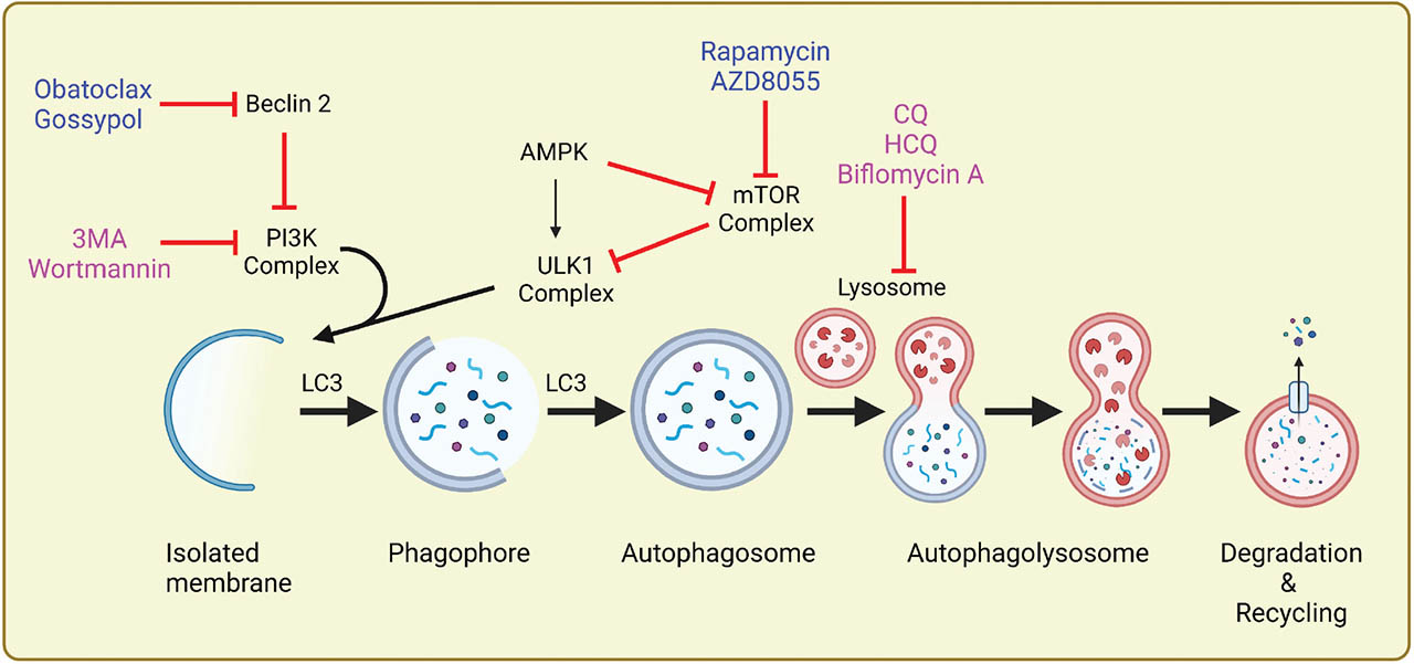
Figure 2
Schematic of macroautophagy flux. Autophagy starts with the development of a phagophore followed by the nucleation and elongation controlled by a class III PI3K complex and subsequently enclosure of cytoplasmic contents or organelles to form an autophagosome. The autophagosome then merges with a lysosome to develop into an autophagolysosome, the ultimate site for degradation and recycling. mTOR negatively controls autophagic flux, and it is inhibited by rapamycin and AZD8055 to induce autophagy. Beclin-2 also negatively controls autophagy by inhibiting the PI3K complex, and obatoclax and gossypol inhibit Beclin-2. 3-Methyladenine and wortmannin are early-stage inhibitors that hinder autophagy by negative control of the class III PI3K, and chloroquine, hydroxychloroquine, and biflomycin A are late-stage inhibitors that block the autophagic flux by interfering with the lysosome directly. Created with BioRender.com. Academic License DS23ZNI6O2. mTOR, mammalian targets of rapamycin; PI3K, phosphatidylinositol 3-kinase; CQ, chloroquine; HCQ, hydroxychloroquine.
Table 1
Autophagy regulators used in cancer therapy
| Autophagy regulator | Type | Mode of action | References | |
|---|---|---|---|---|
| 3-Methyladenine | Inhibitor | P13K inhibitors | Upregulation of p62 protein expression | [60, 70, 71, 72] |
| Wortmannin | Inhibit phagosome formation | [70, 73, 74] | ||
| LY294002 | Promote apoptosis | [71, 75] | ||
| Chloroquine | Lysosome inhibitors | Prevent acidification | [76] | |
| Hydroxychloroquine | Inhibit autophagosome accumulation Inhibit the formation of autolysosome | [77, 78, 79, 80] | ||
| Bafilomycin A | Inhibit autophagic degradation | [81] | ||
| Tioconazol | ATG inhibitors | Inhibit phagophore elongation | [82] | |
| FMK-9a | Inhibit autophagosome fusion | [83] | ||
| Rapamycin | Inducer | mTOR inhibitors | Prevent inactivation of ULK1 phosphorylation | [84] |
| AZD8055 | Activate or induce autophagy Induction of apoptosis Cell cycle arrest | [85, 86] | ||
| Vitamin D | Natural products or mTOR inhibitors | Increase intestinal calcium absorption Induce expression of proapoptotic proteins Induces an autophagic transcriptional signature Induce autophagosome formation | [87, 88] | |
| Resveratrol | Induce apoptotic cell death Suppress growth of cancer cell | [89] | ||
| Curcumin | Induce apoptosis Inhibit phosphorylation of AKT | [90] | ||
| Obatoclax | BH3 Mimetics or Beclin-2 inhibitors | Bcl-2 inhibition | [91, 92] | |
| Gossypol | ROS production Autophagic-mediated necroptosis | [93] | ||
[i] AKT, protein kinase B; mTOR, mammalian targets of rapamycin; ATG, autophagy-related genes; ROS, reactive oxygen species; ULK1, Unc-51 like autophagy activating kinase 1.