
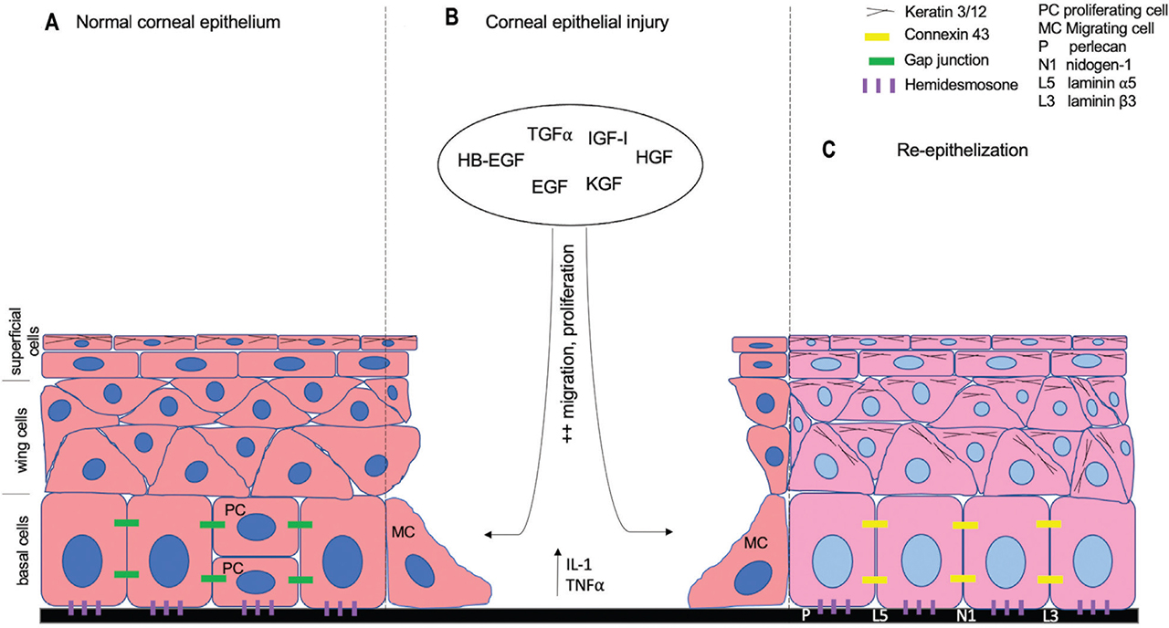
Figure 1
(A) Three main cell types that form the CE. (B) Various growth factors and cytokines are produced in response to injury for the wound healing process. (C) Few markers for nascent regenerating CE and the BM. BM, basement membrane; CE, corneal epithelium.
Table 1
Factors in corneal epithelial wound healing and their functions
| Classification | Factor | Function |
|---|---|---|
| Growth factors | EGF | Promotes cell migration and proliferation |
| Enhances wound closure | ||
| HGF | Enhances cell migration | |
| Upregulates cell proliferation | ||
| Suppresses corneal inflammation | ||
| KGF | Induces corneal epithelial cell proliferation | |
| Enhances stem cells division | ||
| IGF-1 | Has synergistic effect with substance P for epithelial cell migration and cell attachment | |
| IGF-2 | Induces differentiation of limbal stem cells into epithelial cells | |
| Keratin | KRT3/12 or K3/12 | Differentiation marker for terminally differentiated corneal epithelium |
| Structural and mechanical support of the epithelium | ||
| Cytokines | IL-1 | Master regulator in the corneal epithelial wound healing process |
| Accelerates wound healing | ||
| TNF-α | Pro-inflammatory cytokines | |
| Basement membrane proteins | Type IV collagen | Aides cell adhesion, migration and differentiation |
| Laminin | Promotes corneal epithelial cells adhesion | |
| Perlecan | Structural support for epithelial basement membrane | |
| Nidogen | Provide link between components of basement membrane | |
| Gap junction protein | Cx43 | Mediates intercellular communication |
| Integrins | α2, α3, α6, αV, β1, and β4 | Modulate the epithelial shape during migration |
| Ligand for fibronectin, laminin |
[i] Cx, connexin; EGF, epidermal growth factor; HGF, hepatocyte growth factor; IGF-1, insulin growth factor-1; IGF-2, insulin growth factor-2; IL-1, interleukin-1; KGF, keratinocyte growth factor; K3/12, keratin protein 3/12; KRT3/12, keratin gene 3/12; TNF-α, tumor necrosing factor-α