
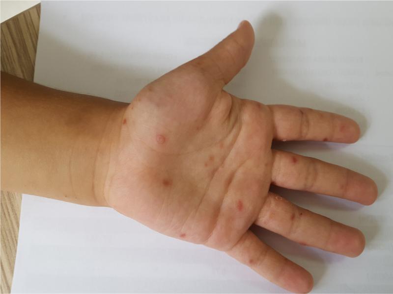
Fig. 1.
Patients with typical lesions on the hands.
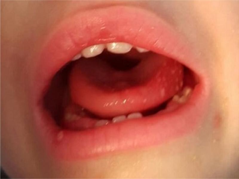
Fig. 2.
Patients with mouth ulcers.
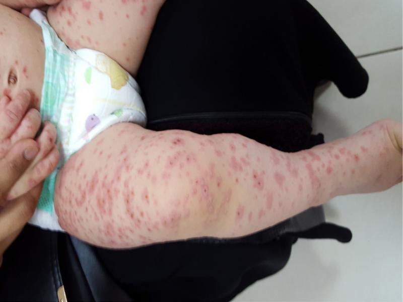
Fig. 3.
Patient with generalized lesions on the legs.
Table I
Primers used for PCR.
| Target Serotype | Orientation | Sequence |
|---|---|---|
| Pan-EV-5’UTR Outer | Forward | 5’-CYTTGTGCGCCTGTTTT-3’ |
| Reverse | 5’-ATTGTCACCATAAGCAGCC-3’ | |
| Pan-EV-5’UTR-Inner | Forward | 5’-CAAGYACTTCTGTMWCCCC-3’ |
| Reverse | 5’-CCCAAAGTAGTCGGTTCC-3’ | |
| EV-A71-VP1 | Forward | 5’-AGAGCATGATTGAGACACG-3’ |
| Reverse | 5’-RTCTTTCTCYTGTTTGTGTTC-3’ | |
| CV-A16-VP1 | Forward | 5’-TGCAGACATGATTGACCAG-3’ |
| Reverse | 5’-TGCCTACAGTTCTGATGCTA-3’ |
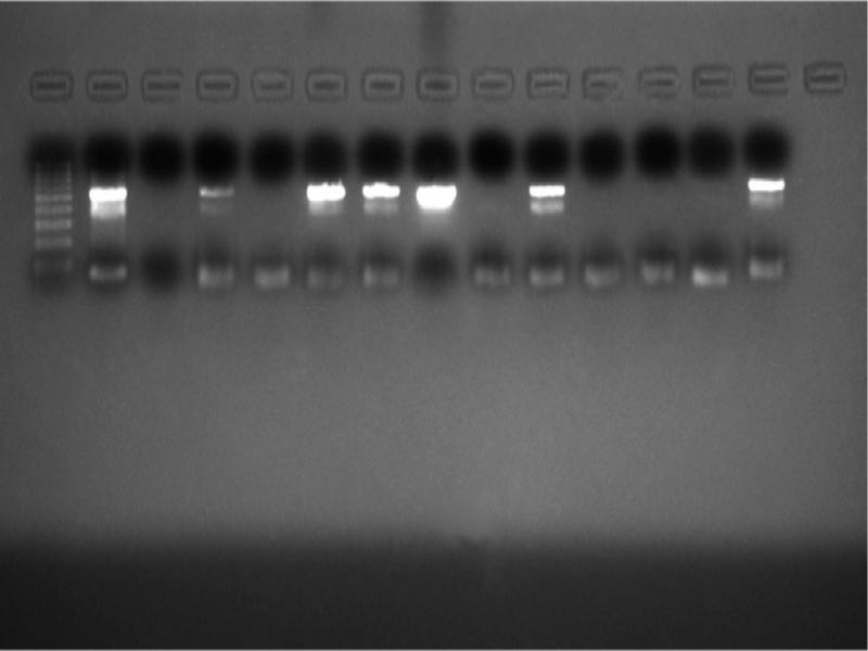
Fig. 4.
The samples were subjected to the pan-enterovirus specific nested PCR with outer primers where 530 bp products were amplified. Left to right, line 1: 100 bp marker, lines 2 to 5: HFM-2 to HFM-5, line 6: HFM-7, line 7: HFM-9, lines 8 to 14: HFM-10 to HFM-16.
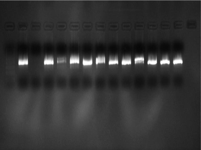
Fig. 5.
The samples were subjected to the pan-enterovirus specific nested PCR with inner primers where 389 bp products were amplified. Left to right, line 1: 100 bp marker, lines 2 to 5: HFM-2 to HFM-5, line 6: HFM-7, line 7: HFM-9, lines 8 to 14: HFM-10 to HFM-16.
Table II
Phylogenetic identities of the coxsackievirus A isolates, based on VP1 sequences.
| Isolate code | Primer | Nearest phylogenetic neighbours | Identity percentage |
|---|---|---|---|
| HFM-2 | 153 F/541 R | Coxsackievirus A6 | 99% |
| HFM-5 | 153 F/541 R | Coxsackievirus A6 | 99% |
| HFM-9 | 153 F/541 R | Coxsackievirus A6 | 99% |
| HFM-10 | 153 F/541 R | Coxsackievirus A6 | 99% |
| HFM-11 | 153 F/541 R | Coxsackievirus A6 | 99% |
| HFM-12 | 153 F/541 R | Coxsackievirus A6 | 99% |
| HFM-14 | 153 F/541 R | Coxsackievirus A6 | 99% |
| HFM-15 | 153 F/541 R | Coxsackievirus A6 | 99% |
| HFM-16 | 153 F/541 R | Coxsackievirus A6 | 99% |
| HFM24 | 153 F/541 R | Coxsackievirus A16 | 99% |
| HFM25 | 153 F/541 R | Coxsackievirus A16 | 99% |
| HFM26 | 153 F/541 R | Coxsackievirus A16 | 97% |
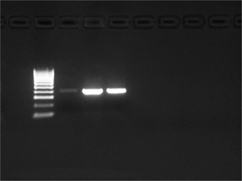
Fig. 6.
CV-A6 specific primers amplified products of approximately 645 bp.
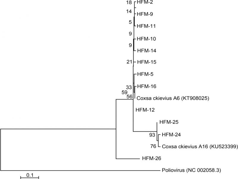
Fig. 7.
Phylogenetic analysis of clinical isolates. Phylogenetic analysis of coxsackievirus A6 nucleotide sequences that were obtained using 153F ve 541R primers, showed the relationships between the clinical CV-A6 and the CV-A16 isolate selected from the GenBank. A scale bar indicates branch distances. The phylogenetic tree was constructed using neighbour-joining method and validated with 1000 pseudo-replicates. Poliovirus (NC 002058.3) was selected as out-group.