
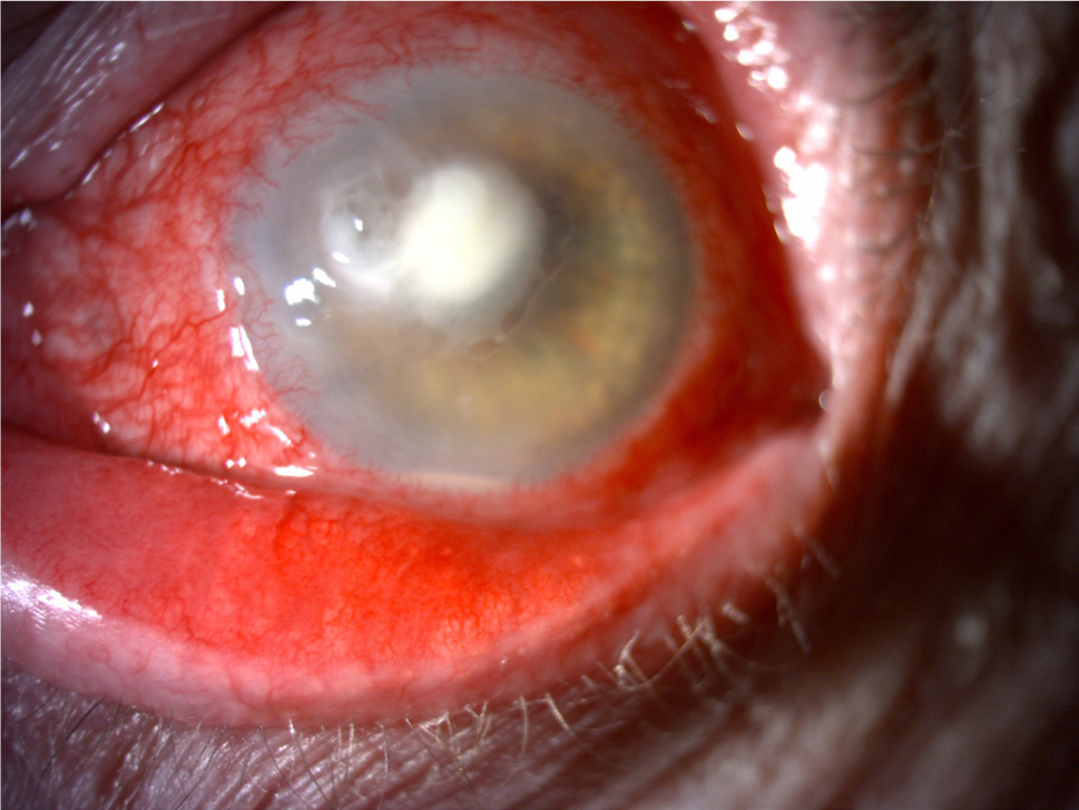
Fig. 1.
The disease symptoms of the cornea from which A. penicillioides was isolated.
Photo E. Machowicz-Matejko.
Table I
Primers used in PCR reaction.
| Name | Primer | Primer sequence 5’-3’ | Melting temperature (Tm) °C | Primer orientation | Reference |
|---|---|---|---|---|---|
| ITS primers | ITS1 | TCCGTAGGTGAACCTGCGG | 59 | Forward | Ref. 28 |
| ITS4 | TCCTCCGCTTATTGATATGC | 59 | Reverse | Ref. 28 | |
| LSU primers | LROR | ACCCGCTGAACTTAAGC | 60 | Forward | Ref. 28 |
| LR5 | TCCTGAGGGAAACTTCG | 60 | Reverse | Ref. 29 | |
| β-tubulin primers | Bt2a | GGTAACCAAATCGGTGCTGCTTTC | 60 | Forward | Ref. 30 |
| Bt2b | ACCCTCAGTGTAGTGACCCTTGGC | 60 | Reverse | Ref. 30 |
Table II
The degree of identity and length of the covered sections of sequences as defined by the BLAST methods.
| Species | Strain/isolate | The analyzed sequences from GenBank accession No. | ||||||||
|---|---|---|---|---|---|---|---|---|---|---|
| ITS | LSU | β-tubulin | ||||||||
| No. GenBank | The degree of identity of sequence [%] = identity | Query cover nucleotides | No. GenBank | The degree of identity of sequence [%] = identity | Query cover nucleotides | No. GenBank | The degree of identity of sequence [%] = identity | Query cover nucleotides | ||
| Aspergillus penicillioides | isolate B1-8 | KF986414.1 | 100 | 569 | ||||||
| Aspergillus penicillioides | UFMGCB 6310 | KF373543.1 | 100 | 498 | ||||||
| Aspergillus sp. | DY115-21-7-M6 | KF411572.1 | 100 | 551 | ||||||
| Uncultured fungus | clone LX042233-122-012-B01 | GQ999240.1 | 99 | 574 | ||||||
| Aspergillus penicillioides | isolate KH00279 | GU017500.1 | 99 | 574 | GU017541.1 | 99 | 835 | |||
| Aspergillus penicillioides | isolate KH00251 | GU017494.1 | 99 | 574 | GU017535.1 | 100 | 833 | |||
| Uncultured fungus | clone PR-MAT-CV5-17 | FJ265955.1 | 99 | 574 | ||||||
| Aspergillus sp. | F55 | FJ755819.1 | 99 | 574 | ||||||
| Aspergillus penicillioides | strain WR1996 | KP997215.1 | 99 | 544 | ||||||
| Aspergillus penicillioides | Culture collection CCF<CZE> 3112 | FR727125.1 | 99 | 573 | ||||||
| Uncultured fungus | clone PR-MAT-CV5-19 | FJ265957.1 | 99 | 571 | ||||||
| Aspergillus penicillioides | isolate HNC15-78 | KT959298.1 | 99 | 530 | ||||||
| Uncultured fungus | clone xnh90 | KP063524.1 | 99 | 574 | ||||||
| Aspergillus sp. | 43m | KC834810.1 | 99 | 527 | ||||||
| Uncultured Aspergillus | clone CHiv38 | KP974191.1 | 99 | 574 | ||||||
| Uncultured fungus | clone xnh20 | KP063454.1 | 99 | 574 | ||||||
| Uncultured Aspergillus | clone Leof80 | KF225869.1 | 99 | 574 | ||||||
| Aspergillus penicillioides | clone KJ34-0.2-39 | KT390118.1 | 99 | 791 | ||||||
| Aspergillus penicillioides | U81265.1 APU81265 | 99 | 783 | |||||||
| Aspergillus sp. | CCF 3112 | FR775323.1 | 97 | 506 | ||||||
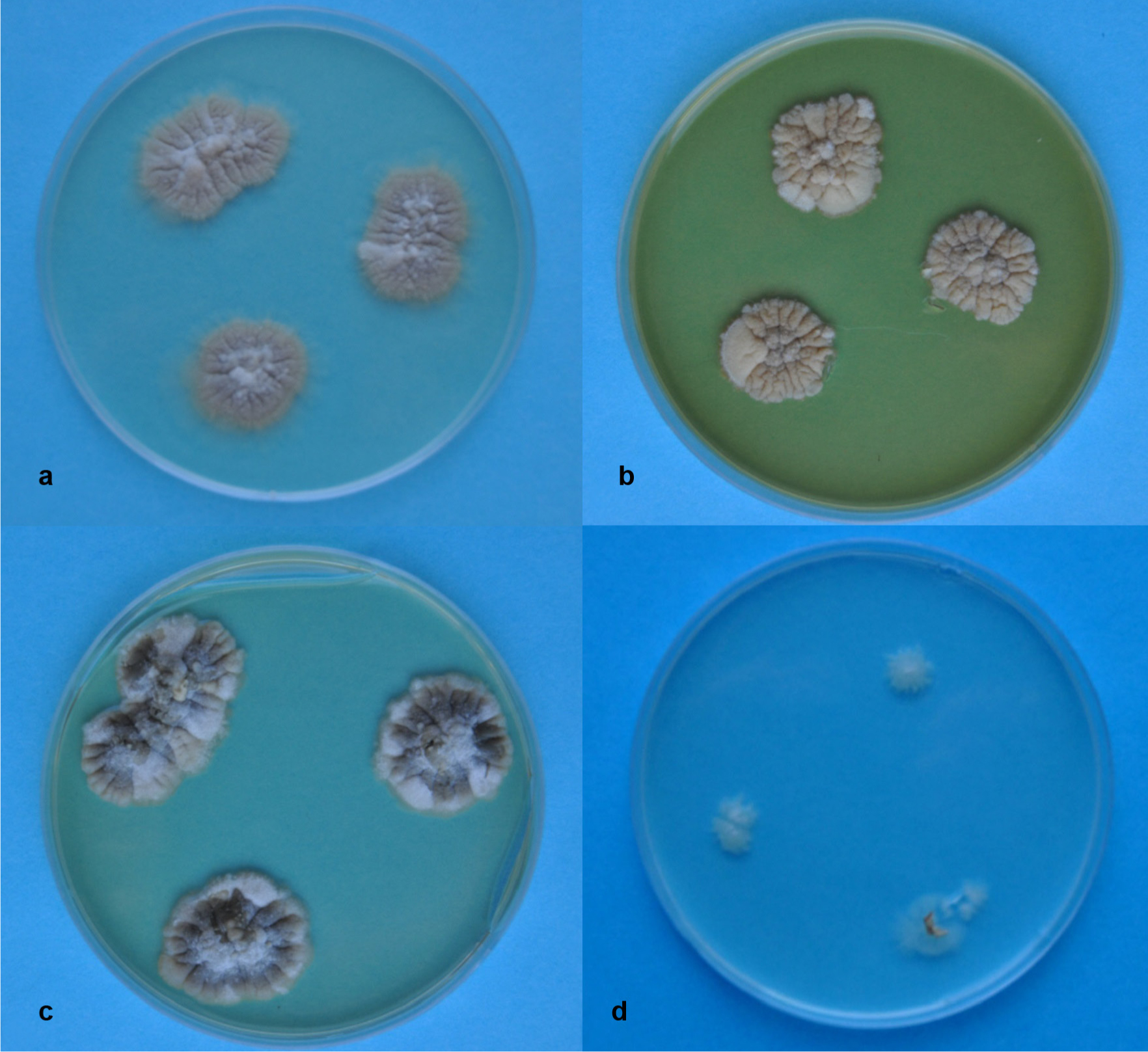
Fig. 2.
Colonies of A. penicillioides after 8 weeks of growing on culture media.
a – PDA, b – YPD, c – Sabouraud, d – a poor medium (see M&M section). Photo E. Zalewska.
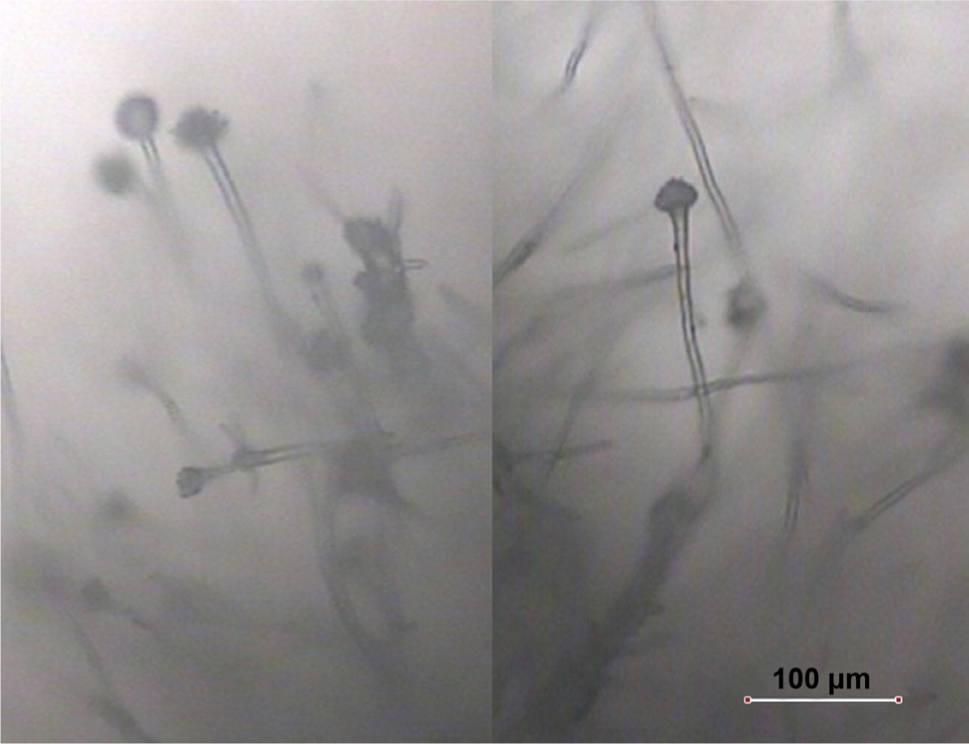
Fig. 3.
A. penicillioides cultured on PDA.
Photo E. Zalewska.
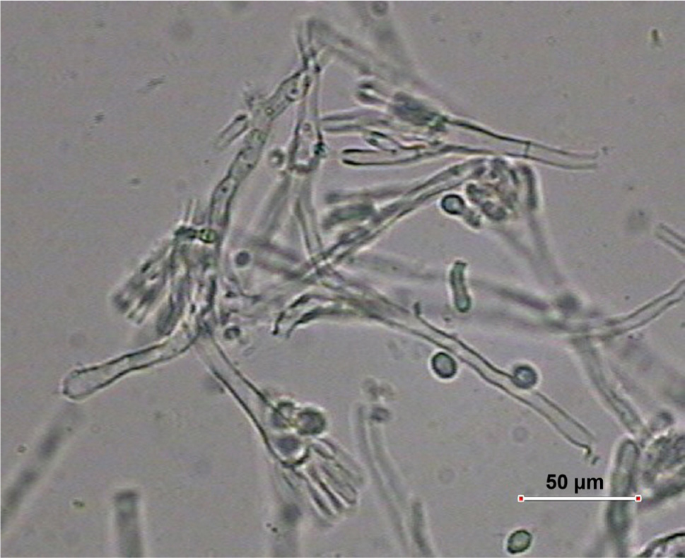
Fig. 4.
Conidiophores and conidia of A. penicillioides on PDA.
Photo E. Zalewska.
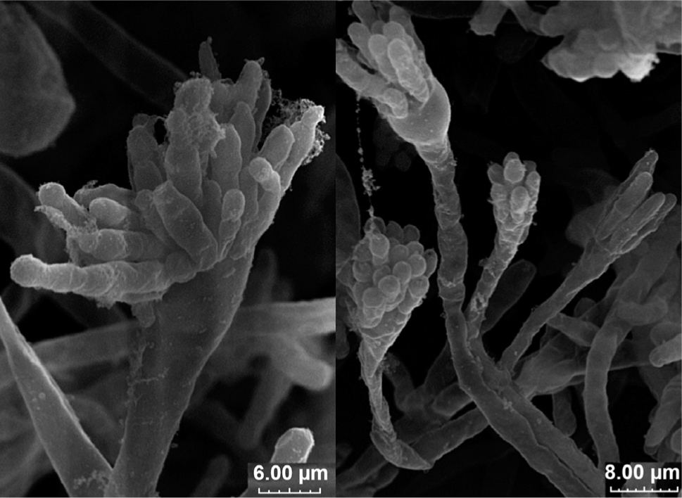
Fig. 5.
Conidiophores of A. penicillioides on PDA.
Photo M. Wróbel.
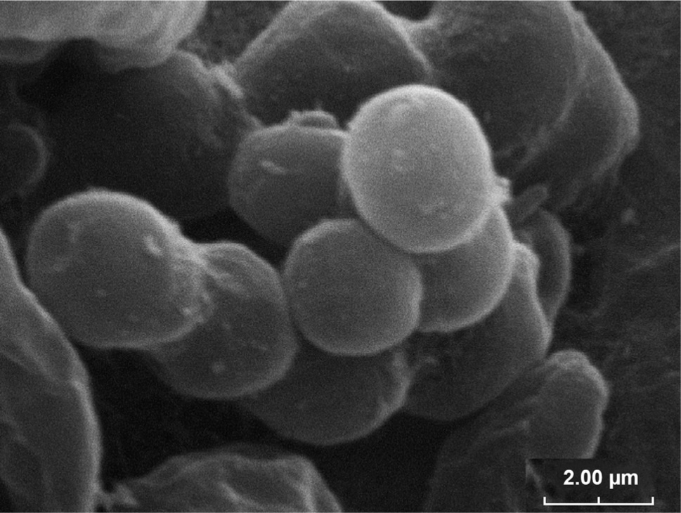
Fig. 6.
Conidia of A. penicillioides on PDA.
Photo M. Wróbel.
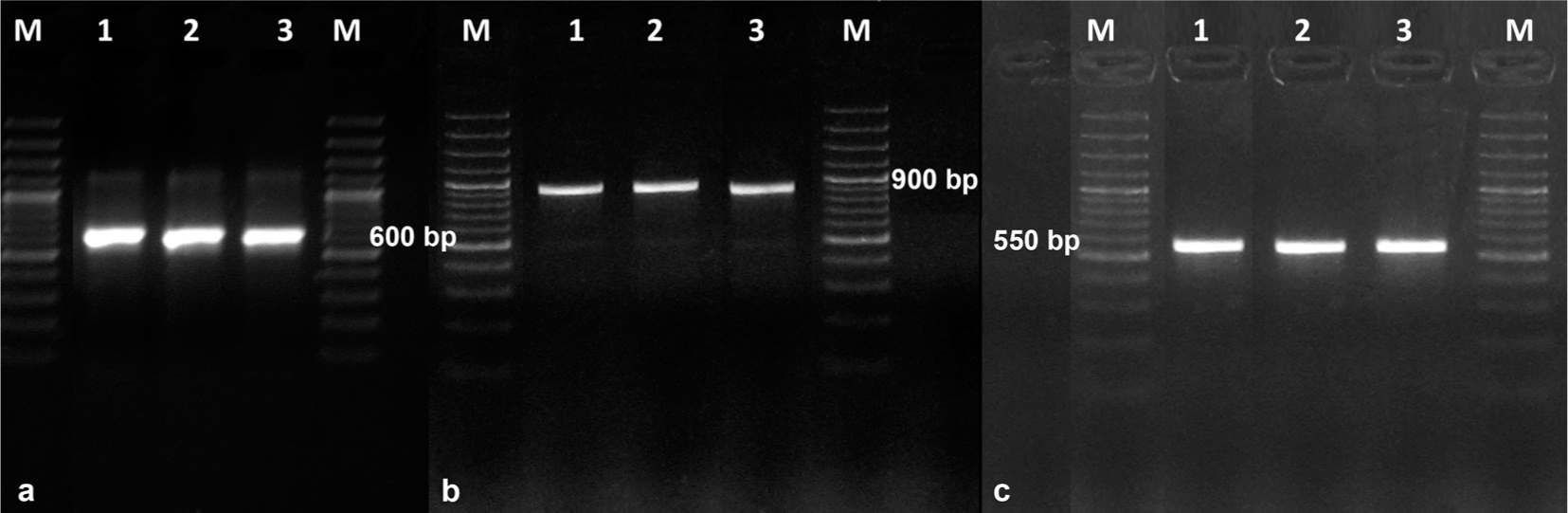
Fig. 7.
Electrophoretic separation of PCR products using universal primers for regions of the genes.
a – rDNA (ITS1-5.8S-ITS4), b – LSU, c – β-tubulin. Photo A. Furmańczyk.
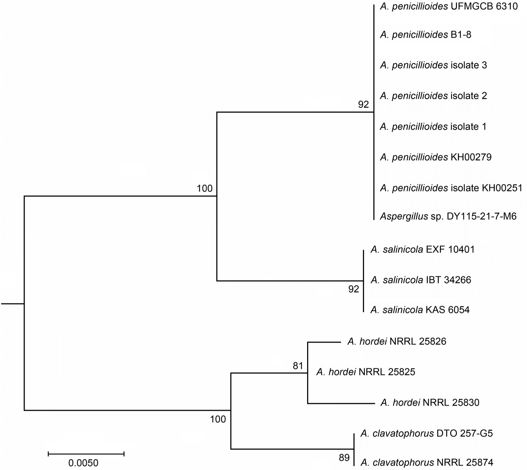
Fig. 8.
Phylogenetic tree of three native A. penicillioides isolates and the reference strains of Aspergillus spp. generated using the Maximum Likelihood method.