
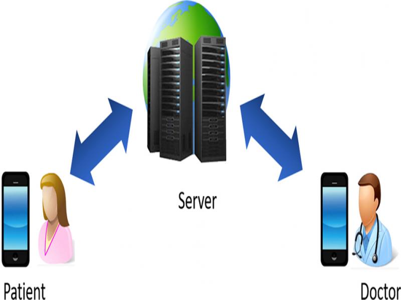
Figure 1:
A telehealth system architecture.
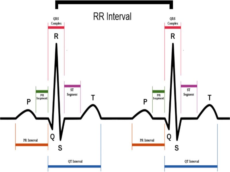
Figure 2:
The illustration of QRS complex in an ECG signal.
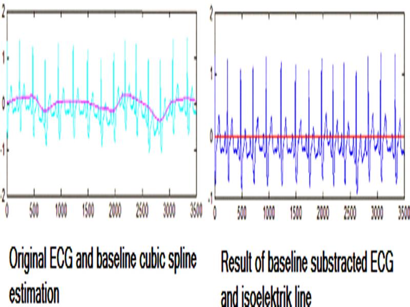
Figure 3:
The ECG data before and after the cubic spline interpolation in order to remove the baseline wander.
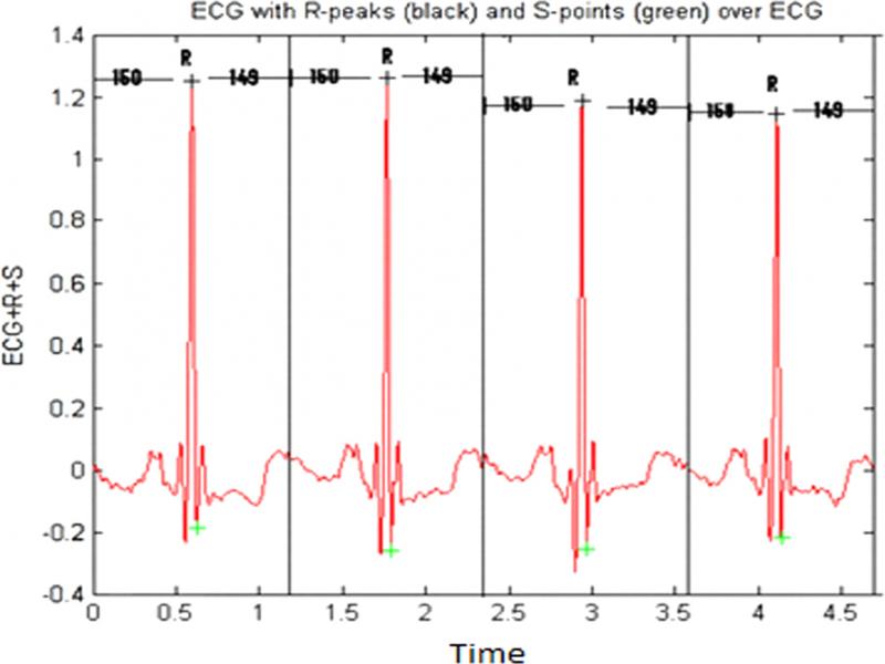
Figure 4:
The individual beat segmentation.
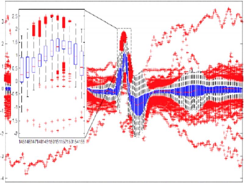
Figure 5:
Outlier removal using IQR.
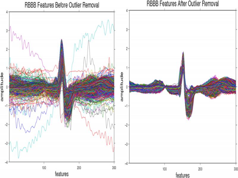
Figure 6:
Beat features before and after outlier removal using IQR.
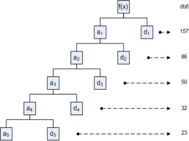
Figure 7:
Daubechies 8 5-level decomposition (Setiawan et al., 2011).
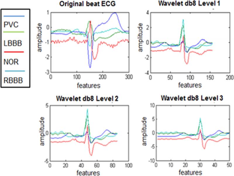
Figure 8:
The ECG decomposed signal.
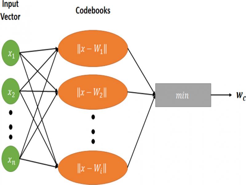
Figure 9:
The architecture of the LVQ algorithm.
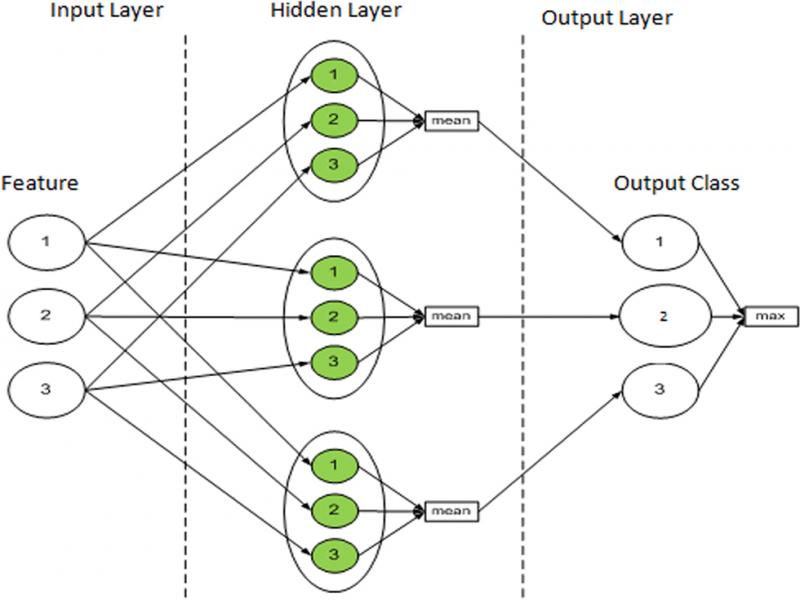
Figure 10:
FNLVQ architecture (Kusumoputro et al., 2002).
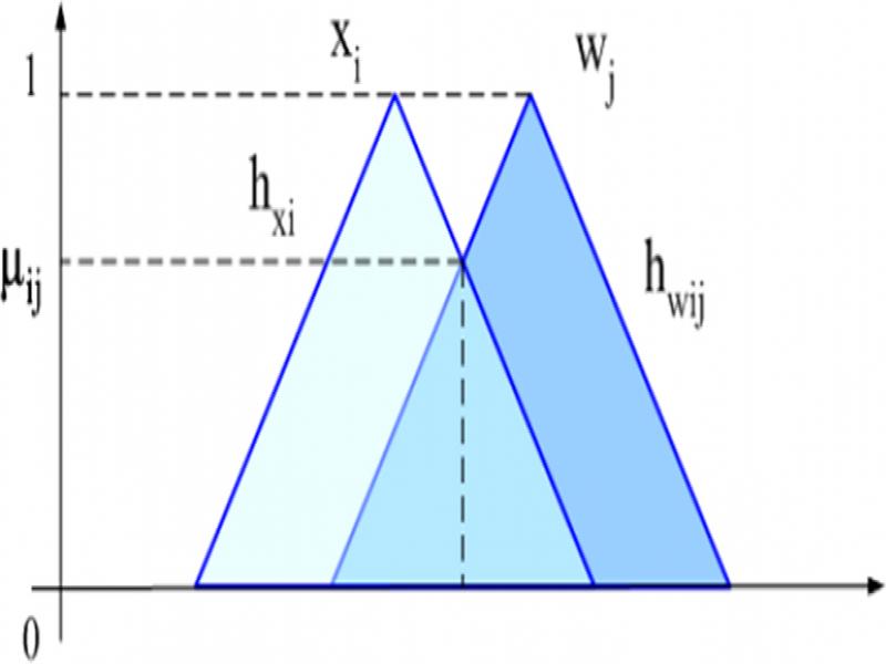
Figure 11:
Computing similarity in FNLVQ (Kusumoputro et al., 2002).
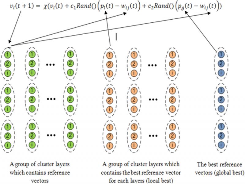
Figure 12:
FNLVQ-PSO architecture (Jatmiko et al., 2009).
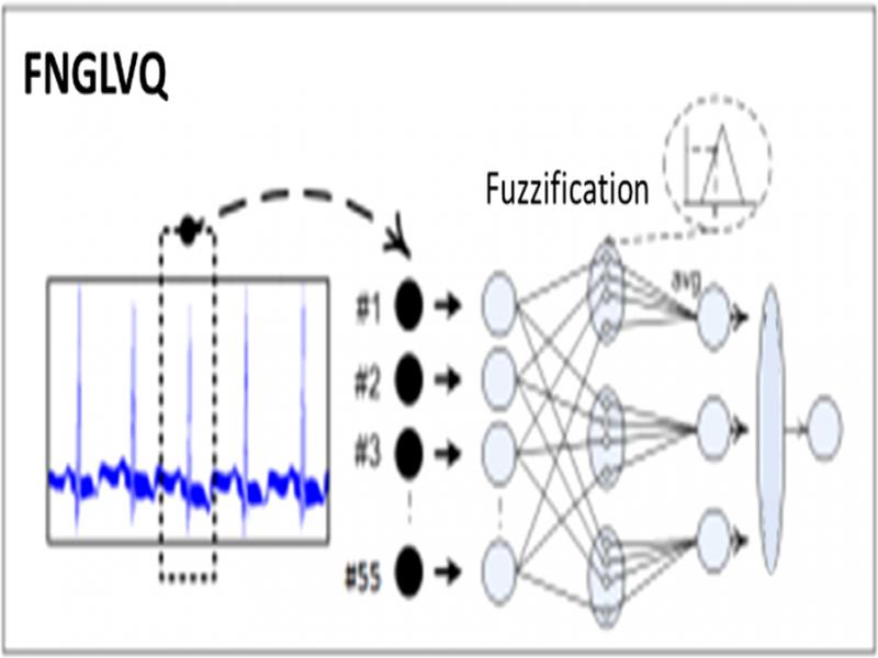
Figure 13:
FNGLVQ architecture (Setiawan et al., 2011).
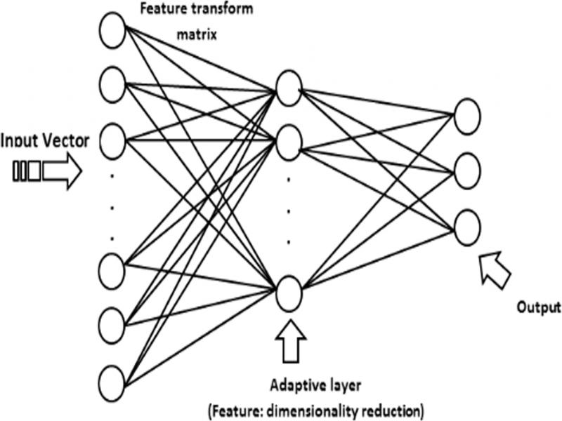
Figure 14:
AM-GLVQ architecture (Imah et al., 2012).
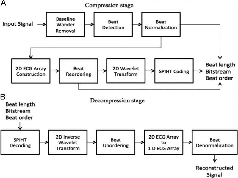
Figure 15:
The process of two-dimensional SPIHT as proposed by Isa et al.
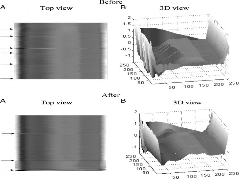
Figure 16:
The ECG data before and after the beat reordering method.
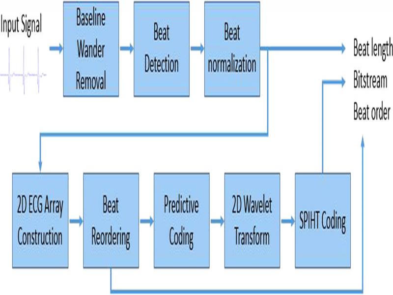
Figure 17:
The process of two-dimensional SPIHT as proposed by Jati et al.
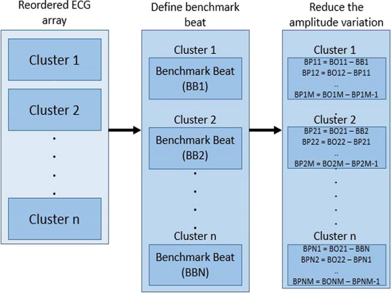
Figure 18:
The process of predictive coding by Jati et al.
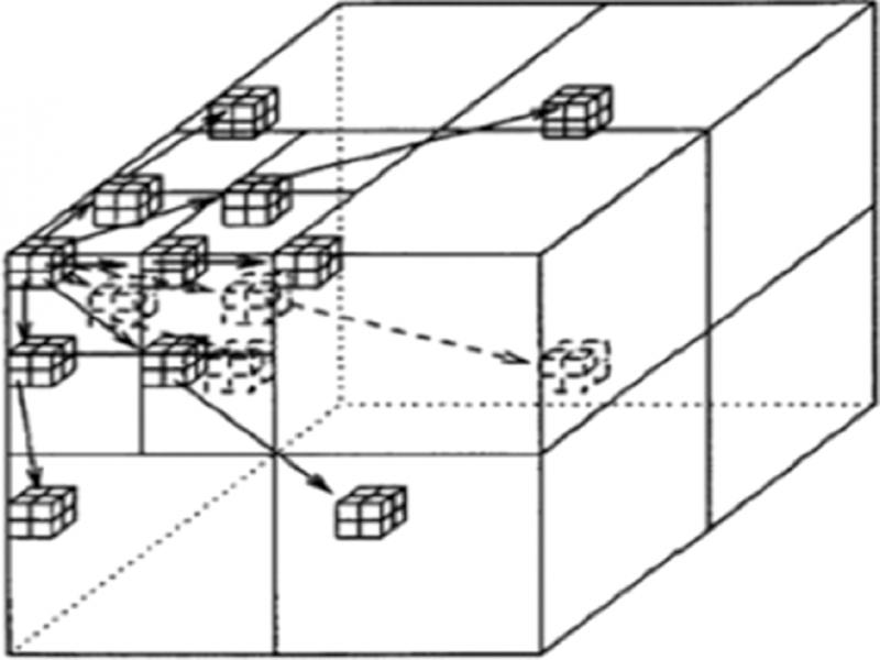
Figure 19:
The spatial orientation tree of 3D SPIHT.
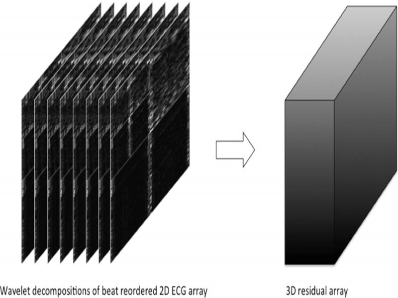
Figure 20:
3D Residual array of the 3D SPIHT method.
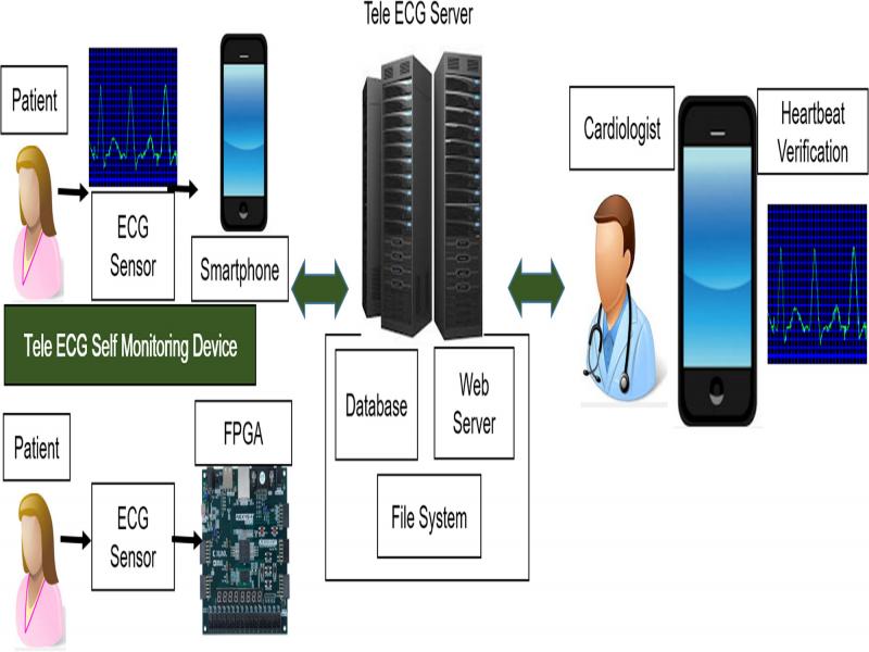
Figure 21:
The architecture of Tele-ECG.
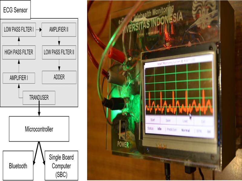
Figure 22:
The ECG sensor.
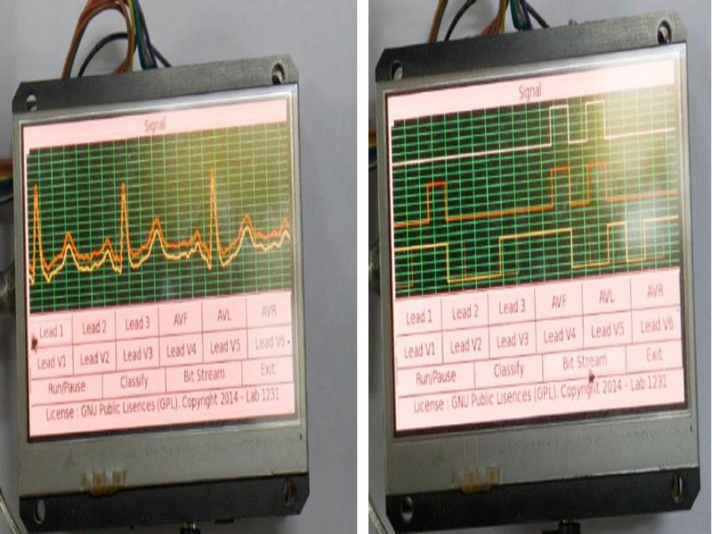
Figure 23:
Multi-lead and single-lead ECG machines.
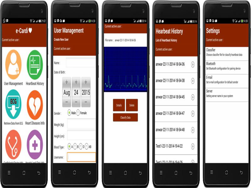
Figure 24:
The mobile system on Tele-EG.
Table 1.
Tele-ECG services.
| No. | URL | Service |
|---|---|---|
| 1 | /RegsiterPatien | Registers user data as patient, then the data are saved in patient table of the database |
| 2 | /RegsiterDoctor | Registers user data as doctor, then the data are saved in doctor table of the database |
| 3 | /UploadHistory | Uploads patient heartbeat history, then the heartbeat data are saved in history table of the |
| 4 | /LookHistory | Checks status of patient heartbeat history if it is verified by a doctor (cardiologist) |
| 5 | /GetDoctorData | Downloads doctor (cardiologists) information |
| 6 | /GetHospitalData | Downloads hospital information |
| 7 | /GetUnverifiedHistory | Downloads unverified patient heartbeat history to be verified by doctor (cardiologist) |
| 8 | /VerifyHistory | Verifies patient heartbeat history by doctor (cardiologist) |
| 9 | /RegisterHospital | Registers hospital data, then the data are saved in hospital table of the database |
| 10 | /RegisterAffiliation | Affiliates the doctor (cardiologist) into hospital |
| 11 | /GetDoctorAffiliation | Downloads doctor’s (cardiologists’) affiliations. |
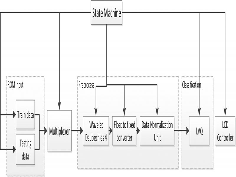
Figure 25:
The ECG classifier on FPGA.
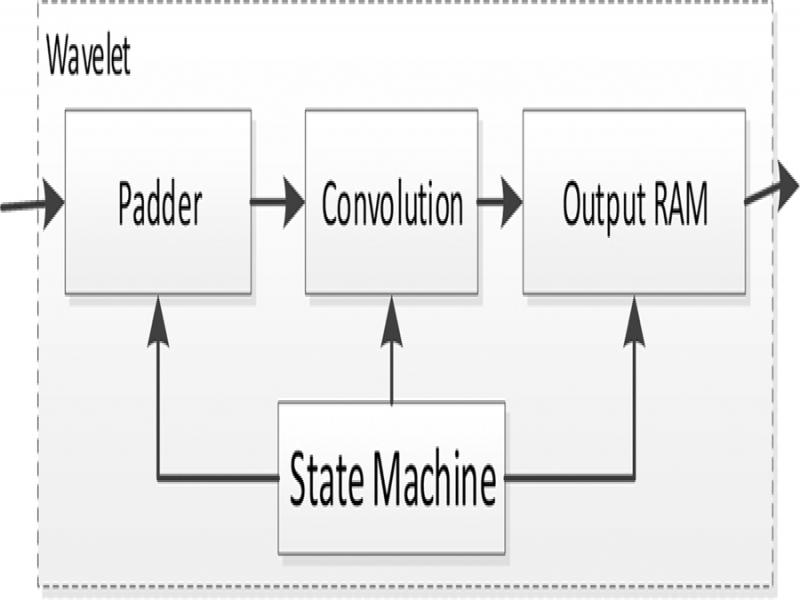
Figure 26:
The Daubechies Wavelet architecture.
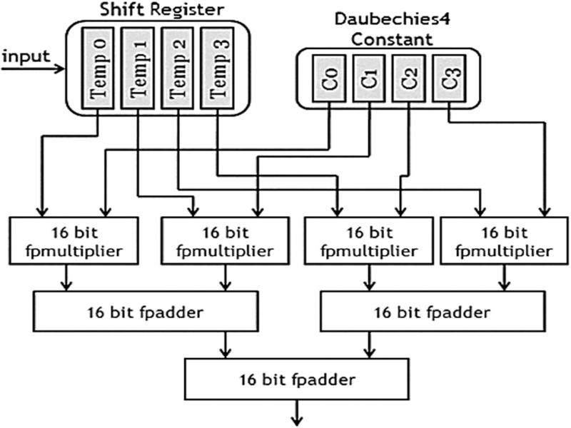
Figure 27:
The Convolution Unit Architecture.
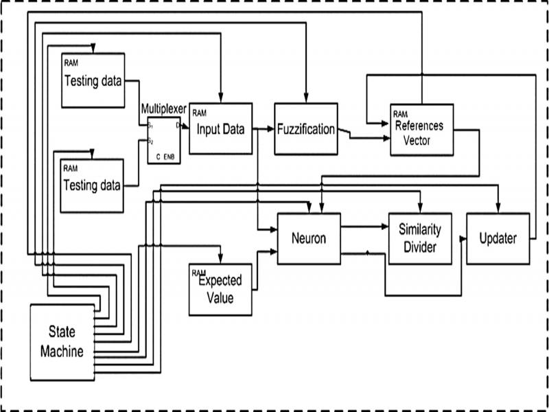
Figure 28:
The design of FLVQ architecture.
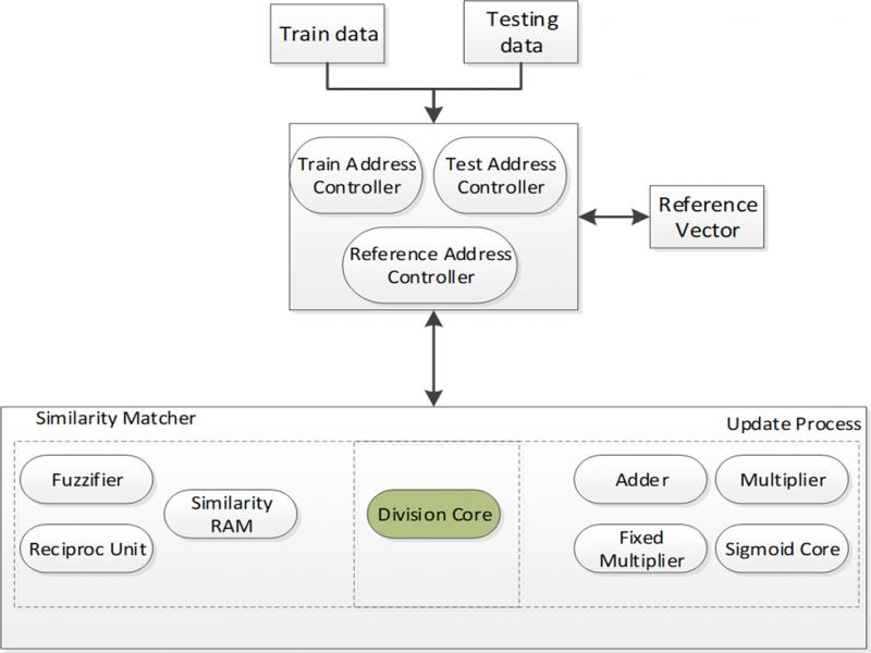
Figure 29:
The design of FNGLVQ in FPGA.
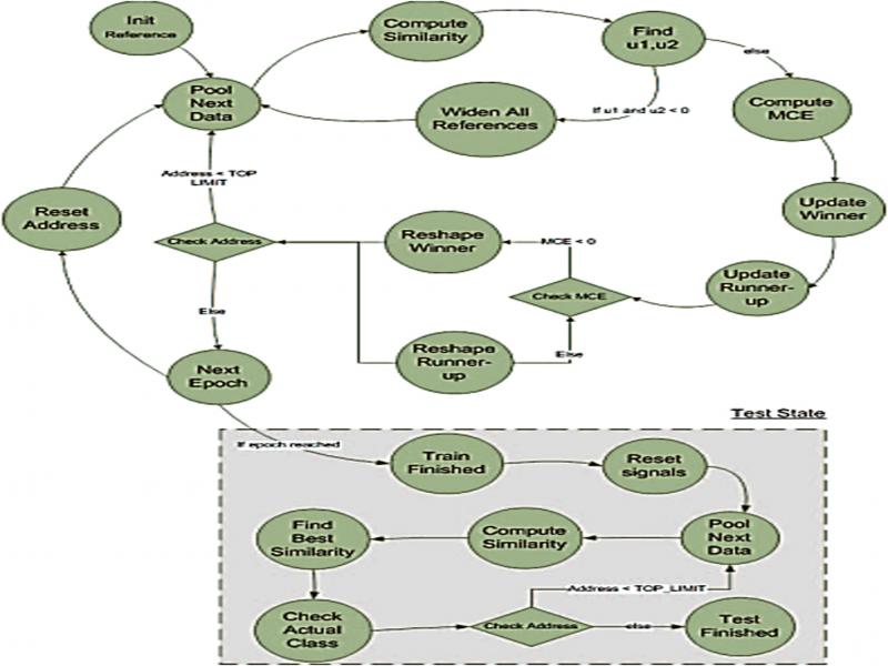
Figure 30:
The State machine of FNGLVQ.
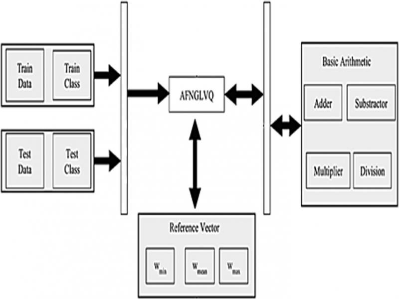
Figure 31:
The top-level design of AFNGLVQ in FPGA.
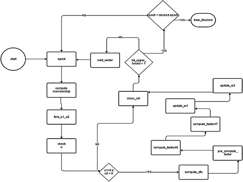
Figure 32:
The top-level design of AFNGLVQ in FPGA.
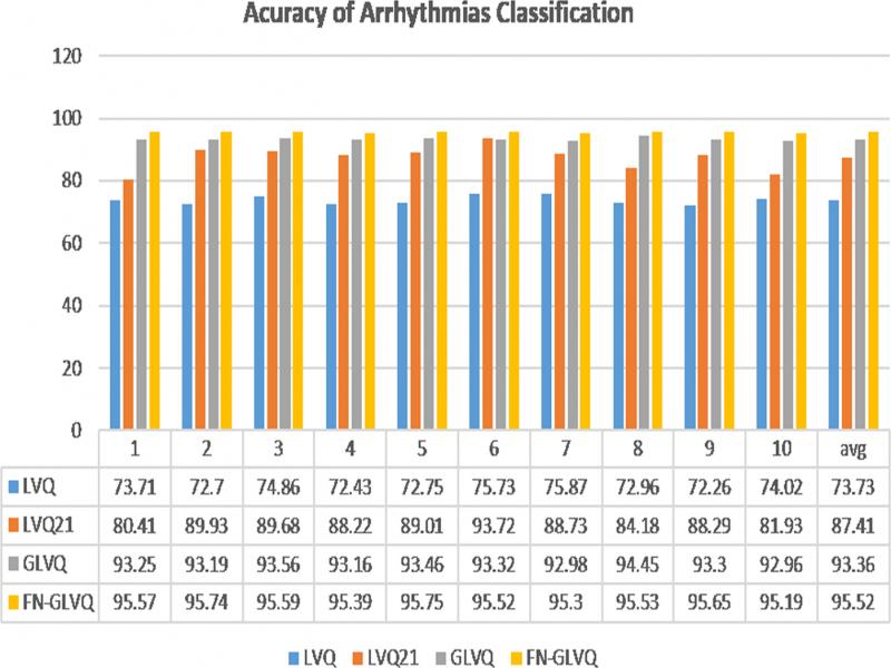
Figure 33:
Accuracy of Arrhythmias classification using LVQ-based classifier.
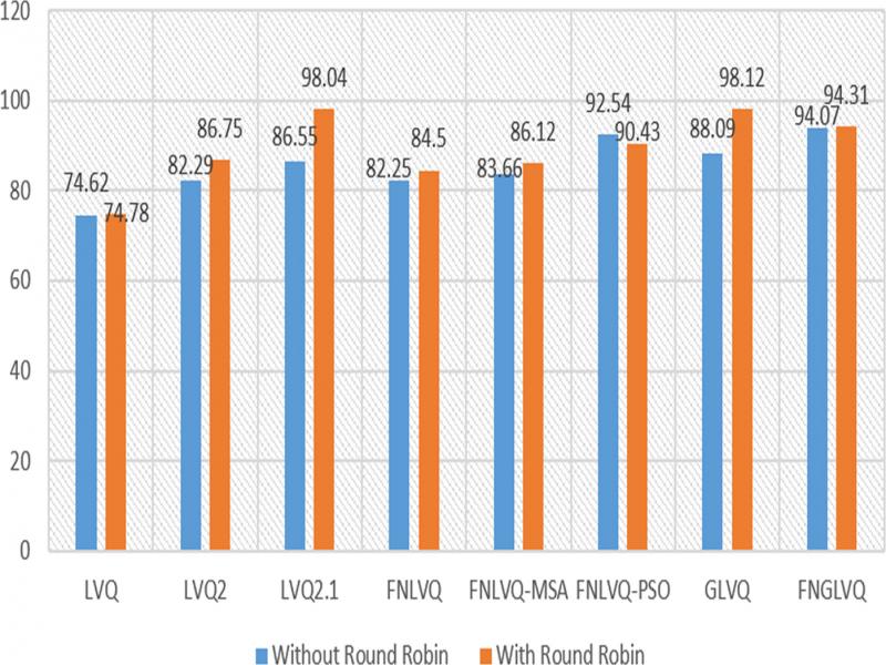
Figure 34:
Impact of Round Robin in Arrhythmias classification.
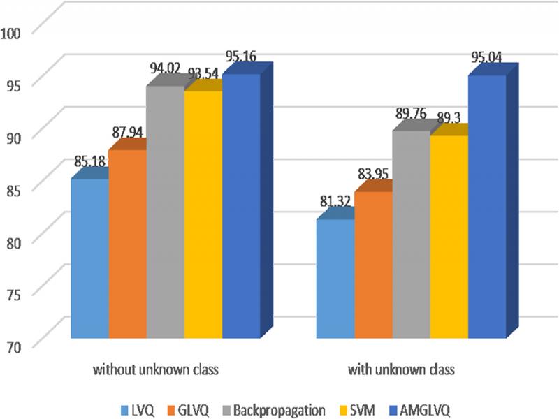
Figure 35:
Arrhythmias classification with unknown class.
Table 2.
ECG compression performance.
| Compression algorithm | Data set | Proposed by | CR | PRD |
|---|---|---|---|---|
| 2D SPIHT | MIT–BIH | Isa et al. (2012b) | 13 | 2.49 |
| 16 | 2.82 | |||
| 3D SPIHT | INCART Arrhythmia | Isa et al. (2014) | 14 | 4.58 |
| 2D SPIHT on embedded device | MIT–BIH | Grafika Jati et al. (2014) | 8 | 1.48 |
| 16 | 2.17 | |||
| 24 | 3.17 | |||
| 32 | 4.06 | |||
| 64 | 11.00 | |||
| 128 | 12.32 |
Table 3.
ECG compression performance.
| No | URL | Mean response time (ms) |
|---|---|---|
| 1 | /RegisterPatient | 129.6 |
| 2 | /RegisterDoctor | 218.9 |
| 3 | /LookHistory | 206.6 |
| 4 | /UploadHistory | 556.4 |
| 5 | /GetHospitalData | 231.7 |
| 6 | /GetDoctorData | 192.3 |
| 7 | /VerifyHistory | 160.7 |
| 8 | /GetUnverifiedHistory | 227.8 |
| 9 | /RegisterAffiliation | 201.8 |
| 10 | /RegisterHospital | 215.0 |
| 11 | /GetDoctorAffiliation | 423.9 |
| Mean | 251.3 | |
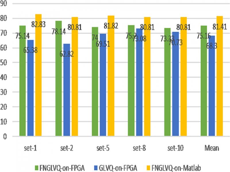
Figure 36:
Arrhythmias classification in FPGA.