
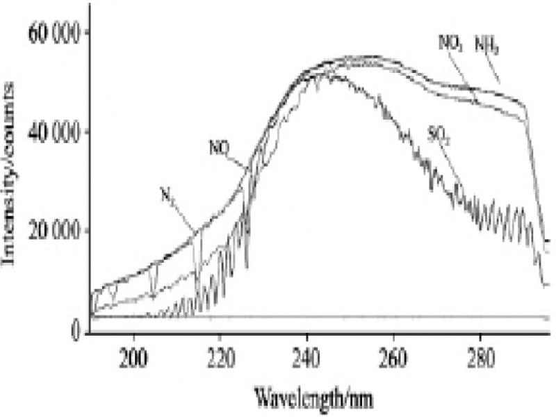
Figure 1.
Mixed gas UV spectral absorption curve
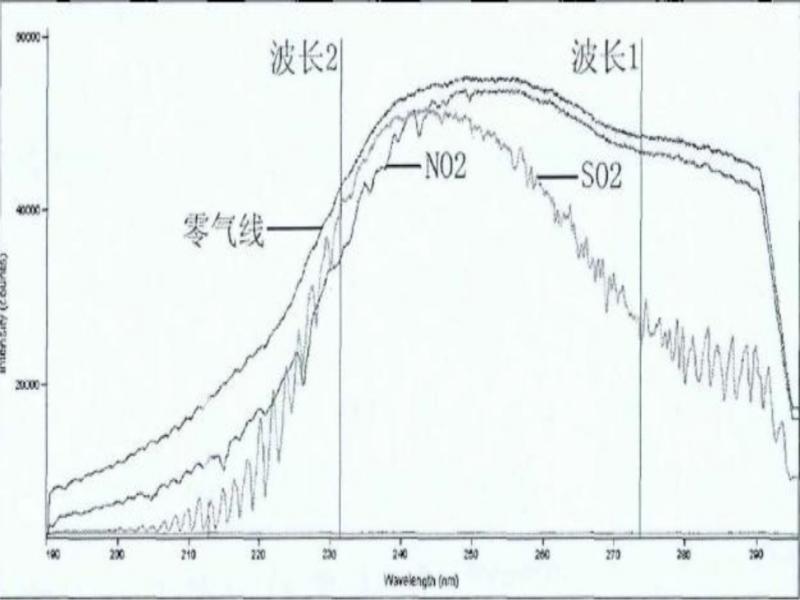
Figure 2.
Absorption spectra of SO2 and NO2
TABLE I.
SPECTRAL TABLES FOR SO2 AND NO2 AT WAVELENGTH 273.33NM AND WAVELENGTH 231.33NM
| Wavelength | 231.33 | 273.33 |
| NO2 absorbance | 0.03003444 | 0.00456189 |
| SO2 absorbance | 0.00482312 | 0.07984884 |
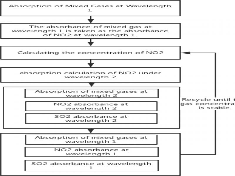
Figure 3.
NO2 and SO2 gas concentration iterative algorithm flow chart
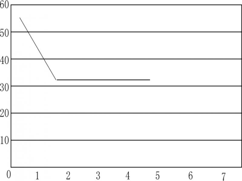
Figure 4.
SO2 concentration and the number of iterations curve
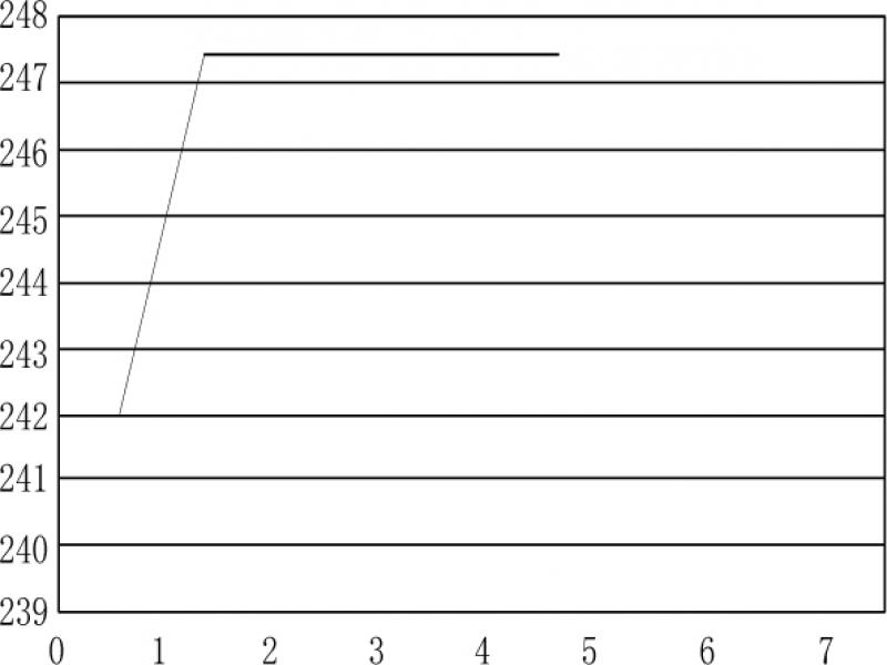
Figure 5.
NO2 concentration and the number of iterations curve
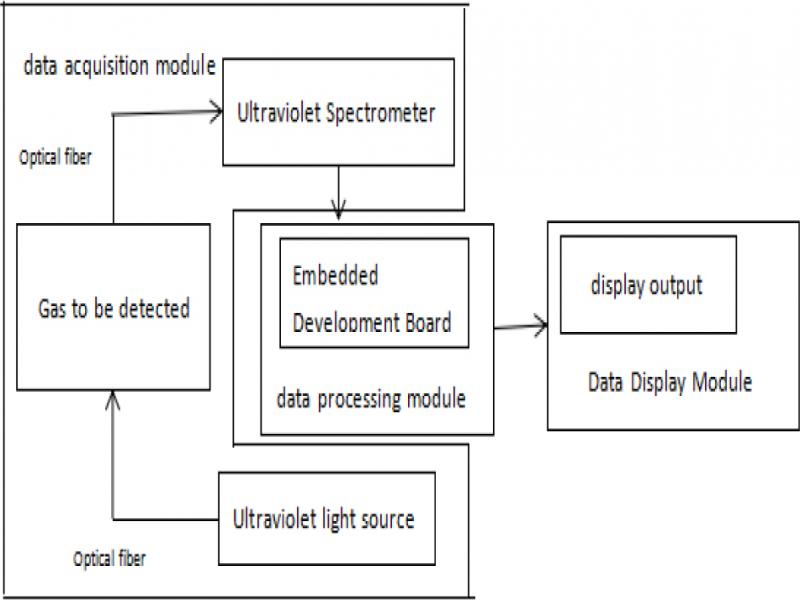
Figure 6.
Experimental system composition diagram
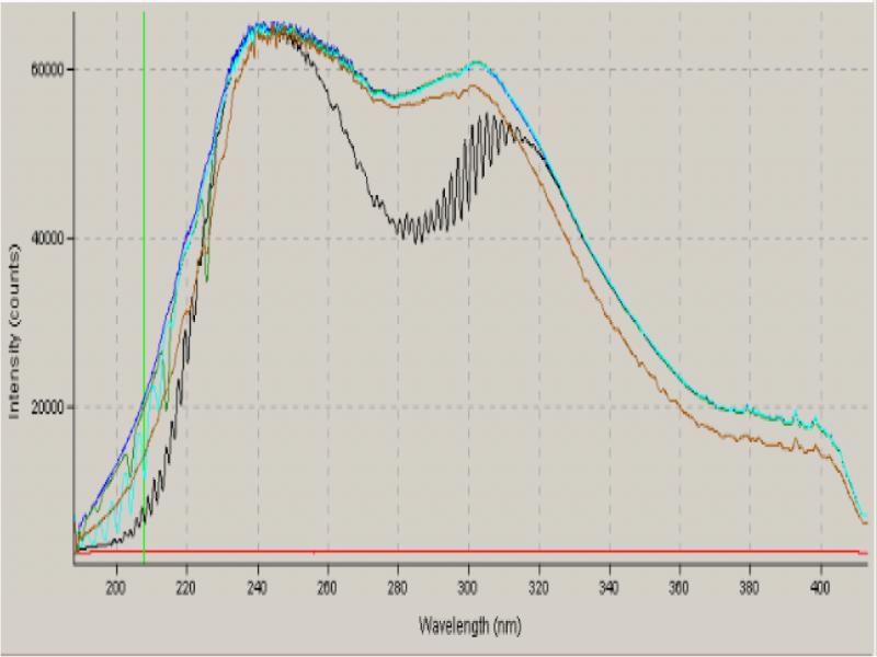
Figure 7.
Four gas photon spectrum line graph
TABLE II.
200PPM SO2 AND NO GAS SPECTRAL DATA
| 271.98nm | 225.94nm | dark noise | |
|---|---|---|---|
| zero gas | 56434 | 43973 | 2900 |
| SO2 100ppm | 52851 | 42754 | 2900 |
| zero gas | 56434 | 43973 | 2900 |
| NO2 100ppm | 56386 | 40749 | 2900 |
TABLE III.
SPECTRAL DATA FOR MIXED GAS
| 271.98nm | 225.94nm | dark noise | |
|---|---|---|---|
| zero gas | 56434 | 43973 | 2900 |
| mixed gas | 52050 | 39588 | 2900 |
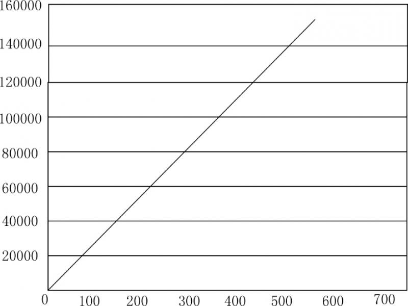
Figure 8.
Fitting curve of SO2 at 271.98 nm
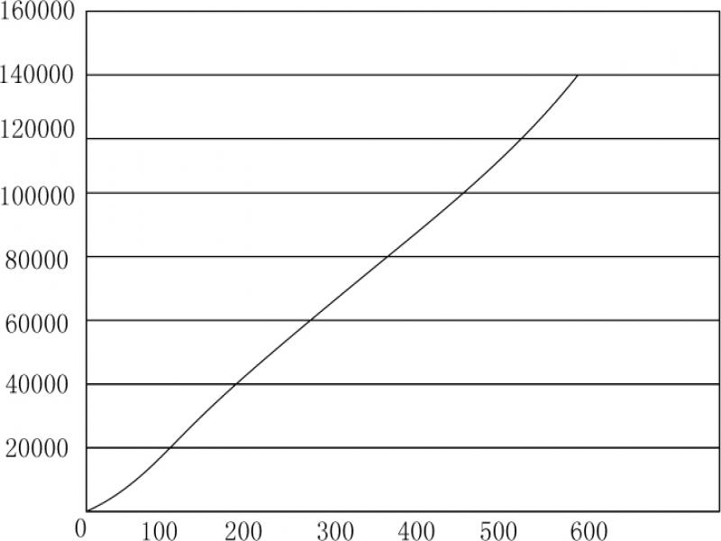
Figure 9.
Fitting curve of NO at 225.94 nm
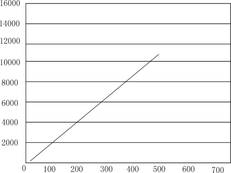
Figure 10.
Fitting curve of NO at 271.98 nm
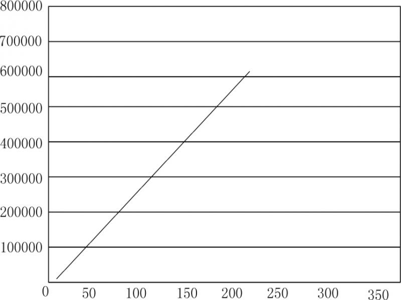
Figure 11.
Fitting curve of NH3 at 271.98 nm
TABLE IV.
ANALYSIS OF RESULTS
| gas species | standard value (ppm) | measured value (ppm) | difference value | maximum difference | error |
|---|---|---|---|---|---|
| SO2 | 102 | 104 | 2 | -4 | 0.8% |
| 200 | 196 | -4 | |||
| 499 | 501 | 2 | |||
| NO | 104 | 108 | 4 | -7 | 1.4% |
| 200 | 206 | 6 | |||
| 500 | 493 | -7 | |||
| NO2 | 116 | 112 | -4 | -9.7 | 1.94% |
| 201.5 | 195 | -6.5 | |||
| 501.7 | 493 | -9.7 | |||
| NH3 | 50 | 53 | 3 | 3 | 1.5% |
| 200 | 199 | -1 | |||
| conclusion | Accuracy error is less than 3%, which meets the design standard. | ||||