
Figure 1.
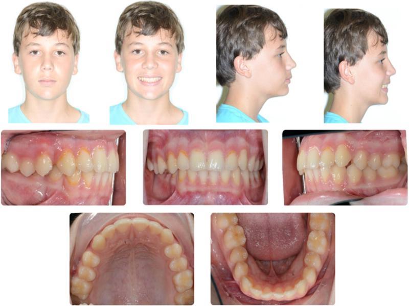
Figure 2.
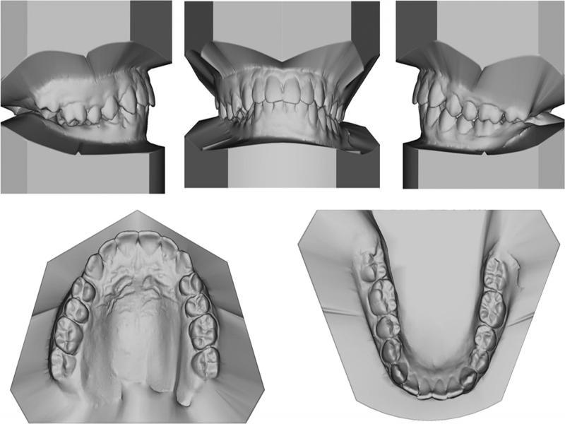
Figure 3.
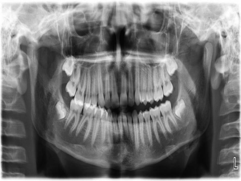
Figure 4.
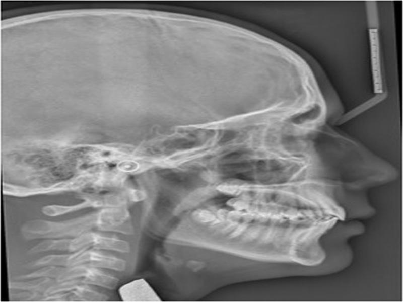
Figure 5.
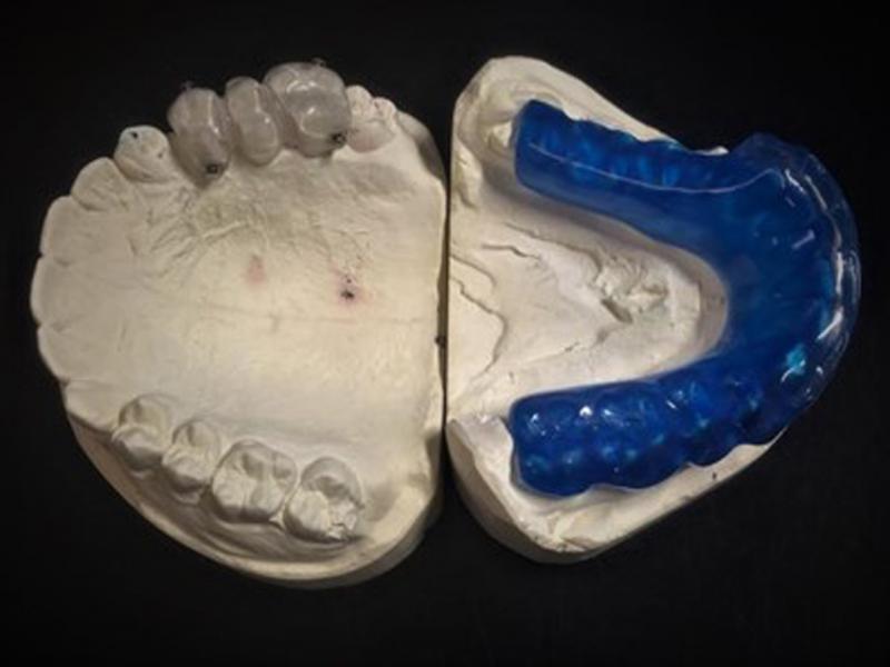
Figure 6.
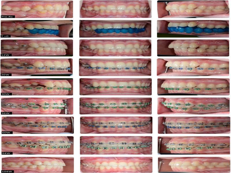
Figure 7.
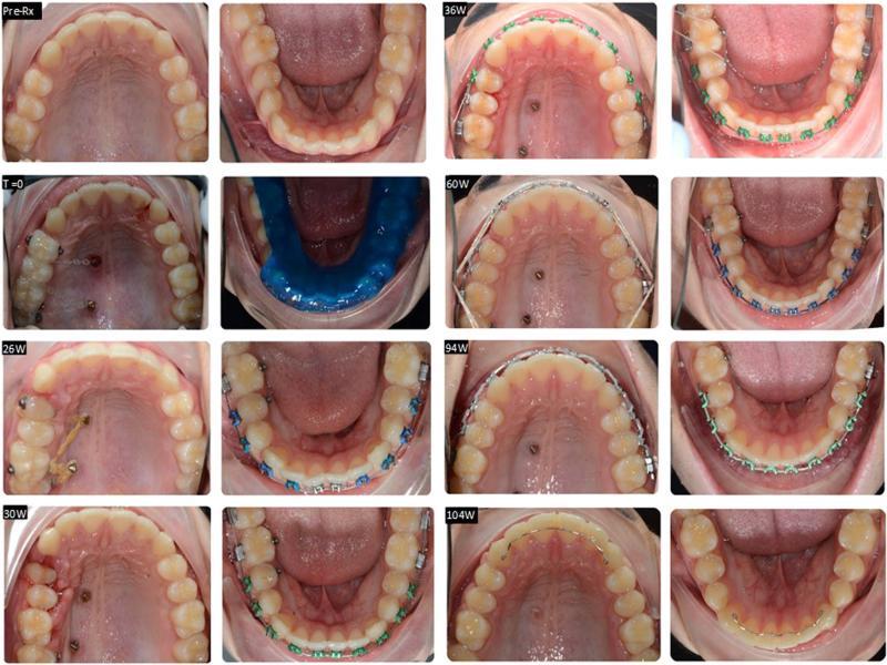
Figure 8.
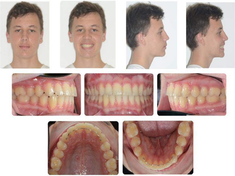
Figure 9.
Figure 10.
Figure 11.
Figure 12.
Figure 13.
Cephalometric summary
| Measurement | Std | Pre-Rx | Post-Rx | Change | |
|---|---|---|---|---|---|
| SKELETAL | |||||
| Maxilla | SNA | 82 | 88 | 88.3 | 0.3 |
| A Point Convexity | 3 | 2.9 | 2.5 | -0.4 | |
| NaVert - A point | 1 mm | 1.8 | 2.6 | 0.8 | |
| Mandible | SNB | 80 | 83.3 | 84 | 0.7 |
| Facial Angle | 87 | 88.9 | 92.2 | 3.3 | |
| NaVert - Po | -8 to -6 mm/-2 to 4 mm | -2.1 | 2.1 | 4.2 | |
| Basal Arch Relationship | ANB | 2 | 4.7 | 4.3 | -0.4 |
| WITS | F: 0 mm | 3.1 | 0 | -3.1 | |
| Vertical | Md Plane Angle | 26 | 20.9 | 18.7 | -2.2 |
| LFH Angle | 47 | 43.9 | 46.3 | 2.4 | |
| Md Arc | 26 | 37.1 | 39.8 | 2.7 | |
| Facial Axis Angle | 90 | 90.7 | 90.8 | 0.1 | |
| Jarabak ratio | 0.59.0.63 | 0.73 | 0.74 | 0.01 | |
| DENTAL | |||||
| Upper Incisors | UI to FH | 110° | 115.9 | 120.4 | 4.5 |
| UI to Pal Plane | 110° | 114.7 | 117.5 | 3.8 | |
| UI to Na Vert | 5 mm | 3.3 | 5 | 1.7 | |
| Lower Incisors | LI to Md Plane | 92.5° | 101.6 | 104.5 | 2.9 |
| LI to Apo | 22 | 28.2 | 31.7 | 3.5 | |
| LI to Apo | 1 | 2.7 | 4.5 | 1.8 | |
| Interincisal | UI to LI | 130 | 121.6 | 114.4 | -7.2 |
| SOFT TISSUE | L Lip - E Line | -2 | -2.4 | -1.1 | 1.3 |
| SnVert - U Lip | 1 mm | 2.2 | 3.3 | 1.1 | |
| SnVert - L Lip | -1 mm | -4.1 | 0.2 | 4.3 | |
| SnVert - Po' | -3 mm | -8.4 | -3.8 | 4.6 | |