
Figure 1.
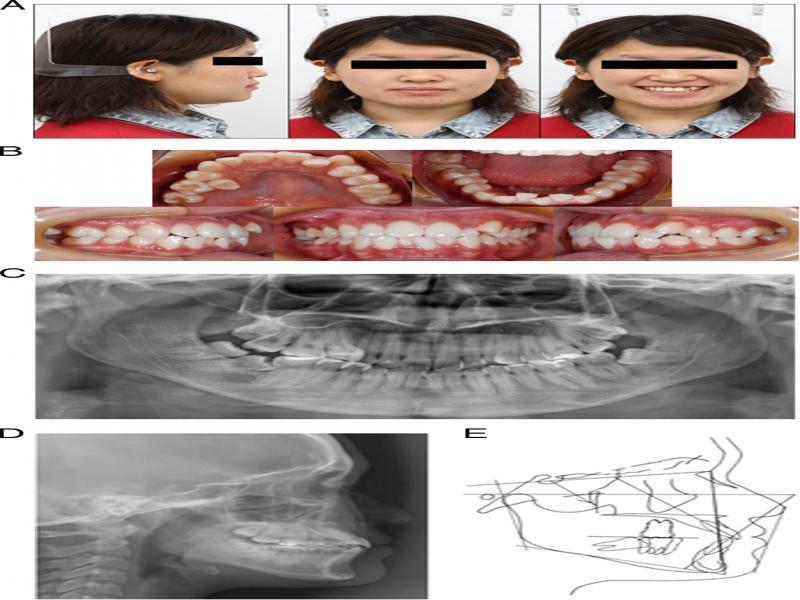
Figure 2.
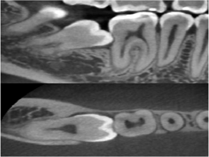
Figure 3.

Figure 4.
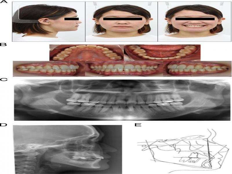
Figure 5.
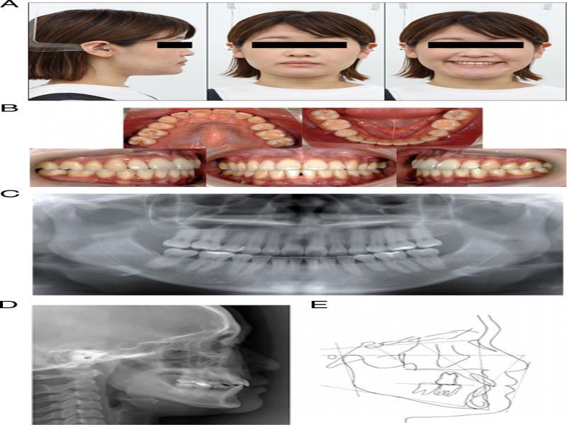
Figure 6.
Cephalometric analyses
| Pretreatment | Posttreatment | After retention | Normative mean | |
|---|---|---|---|---|
| SNA (º) | 82.0 | 82.0 | 82.0 | 82.1 |
| SNB (º) | 82.0 | 82.0 | 82.0 | 78.5 |
| ANB (º) | 0.0 | 0.0 | 0.0 | 3.6 |
| Facial angle (º) | 92.5 | 93.0 | 93.0 | 84.6 |
| Y-axis (º) | 58.0 | 58.0 | 58.0 | 65.2 |
| FMA (º) | 22.5 | 22.0 | 22.0 | 28.6 |
| SN-MP (º) | 32.5 | 32.0 | 32.0 | 34.7 |
| Gonial angle (º) | 121.0 | 121.0 | 121.0 | 121.2 |
| Occ. Plane to SN (º) | 12.0 | 13.5 | 13.5 | 16.8 |
| U1 to SN (º) | 114.0 | 111.0 | 111.0 | 103.8 |
| IMPA (L1 to MP) (º) | 94.0 | 90.0 | 90.0 | 96.2 |
| Interincisal angle (º) | 119.5 | 127.0 | 127.0 | 125.6 |
| U1 to A-Pog (mm) | 8.0 | 6.0 | 6.0 | 7.1 |
| L1 to A-Pog (mm) | 5.0 | 3.0 | 3.0 | 3.6 |
| E-line:Upper (mm) | -3.5 | -4.0 | -2.0 | 0.2 |
| E-line:Lower (mm) | -1.0 | -4.0 | -1.0 | 2.1 |
| Overjet (mm) | R + 4.0, L + 2.0 | 3.0 | 3.0 | 3.1 |
| Overbite (mm) | R + 3.0, L + 1.5 | 2.0 | 2.0 | 3.3 |