
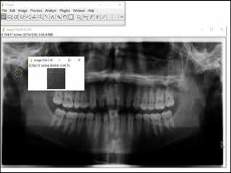
Figure 1.
Panoramic radiograph with selected and duplicated regions of interest (ROI).
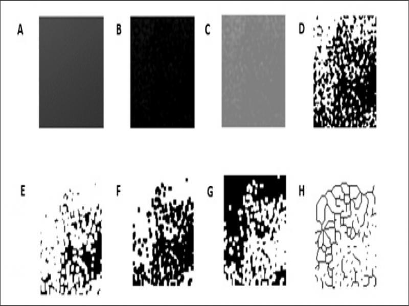
Figure 2.
A, Filter of Gaussian blur. B, Substracted image. C, Addition of 128 gray value. D, Binarisation. E, Erosion. F, Dilatation. G, Inversion. H, Skeletonisation.
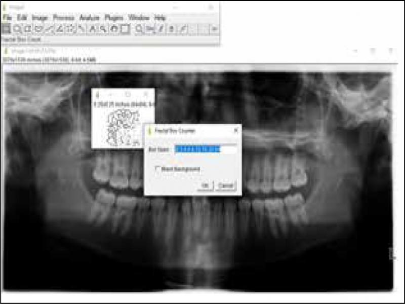
Figure 3.
Fractal box-counting function.
Table I.
Intragroup comparison of fractal dimensions between right and left condyles in skeletal Class I, II div 1, II div 2 and III groups using Student’s t-test.
| Group | FD (Mean ± SD) | p | |
|---|---|---|---|
| Right TMJ | Left TMJ | ||
| Class I | 1.405 ± 0.084 | 1.385 ± 0.090 | 0.257 |
| Class II division 1 | 1.350 ± 0.092 | 1.333 ± 0.088 | 0.362 |
| Class II division 2 | 1.375 ± 0.109 | 1.346 ± 0.100 | 0.174 |
| Class III | 1.334 ± 0.105 | 1.324 ± 0.090 | 0.609 |
Table II.
Intergroup comparison of fractal dimensions using One-way ANOVA and post-hoc Tukey tests.
| Test | Right TMJ | Left TMJ | ||
|---|---|---|---|---|
| One-way ANOVA (p value) | 0.002 | 0.007 | ||
| Pairwise comparison by Tukey test (p value) | I-III | 0.002 | I-III | 0.005 |
| I-II.1 | 0.026 | I-II.1 | 0.027 | |
| I-II.2 | 0.421 | I-II.2 | 0.155 | |
| II.2-III | 0.157 | II.2-III | 0.623 | |
| II.1-II.2 | 0.577 | II.1-II.2 | 0.900 | |
| II.1-III | 0.850 | II.1-III | 0.956 | |
Table III.
Intragroup comparison of fractal dimensions between male and female patients using Student’s t-test.
| Group | Side | FD (Mean ± SD) | p | |
|---|---|---|---|---|
| Male | Female | |||
| Class I | Right | 1.391 ± 0.076 | 1.419 ± 0.091 | 0.251 |
| Left | 1.368 ± 0.091 | 1.401 ± 0.087 | 0.201 | |
| Class II division 1 | Right | 1.358 ± 0.095 | 1.342 ± 0.089 | 0.543 |
| Left | 1.368 ± 0.072 | 1.298 ± 0.089 | 0.004 | |
| Class II division 2 | Right | 1.394 ± 0.104 | 1.354 ± 0.107 | 0.181 |
| Left | 1.348 ± 0.087 | 1.344 ± 0.108 | 0.869 | |
| Class III | Right | 1.356 ± 0.097 | 1.312 ± 0.110 | 0.141 |
| Left | 1.346 ± 0.090 | 1.302 ± 0.087 | 0.083 | |