
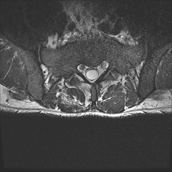
Figure 1a
MRI Without Contrast Axial.
Note. T2-weighted MRI without contrast showing displacement of nerve roots to the periphery of the thecal sac at the levels of L4–S1, known as ‘empty thecal sac sign,’ and is indicative of adhesive arachnoiditis.
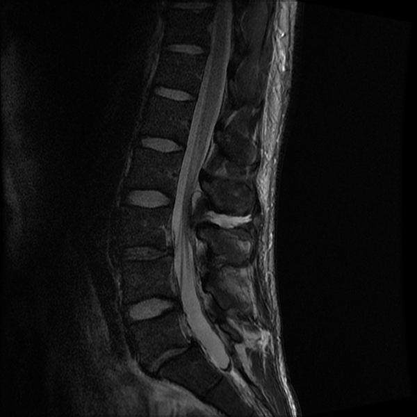
Figure 1b
MRI Without Contrast Sagittal.
Note. T2-weighted MRI without contrast showing displacement of nerve roots to the periphery of the thecal sac at the levels of L4–S1, known as ‘empty thecal sac sign,’ and is indicative of adhesive arachnoiditis.
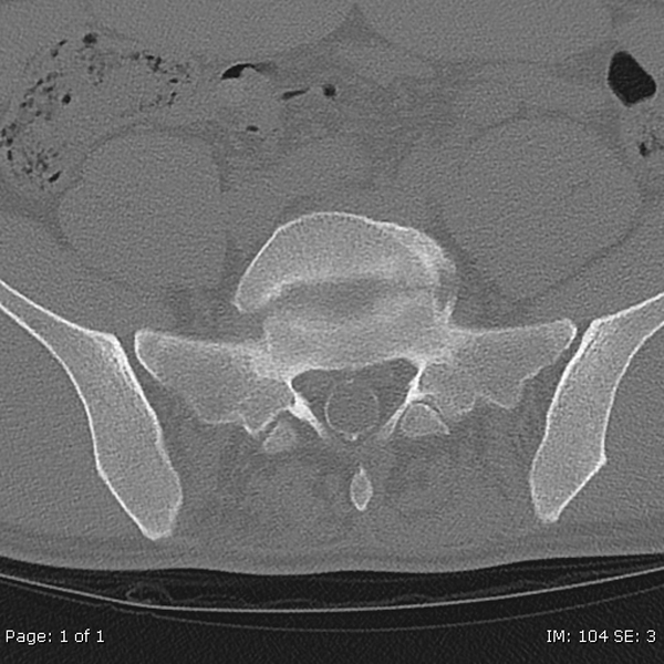
Figure 2a
CT Without Contrast Axial.
Note. CT without contrast demonstrates calcifications along the dura and thecal sac extending from L3 into the sacrum.
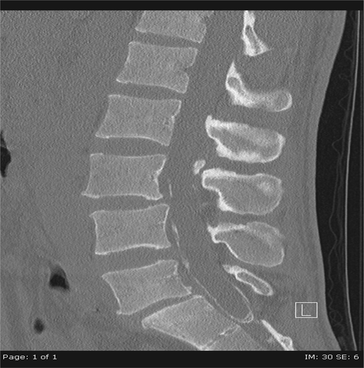
Figure 2b
CT Without Contrast Sagittal.
Note. CT without contrast demonstrates calcifications along the dura and thecal sac extending from L3 into the sacrum.