
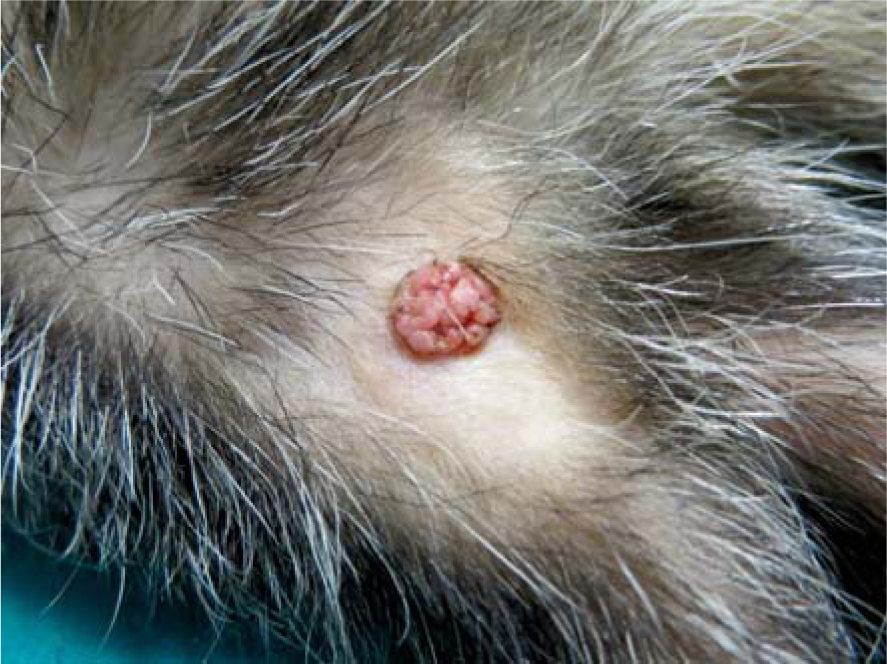
Figure 1
Macroscopic presentation of squamous papilloma (Tumour 4).
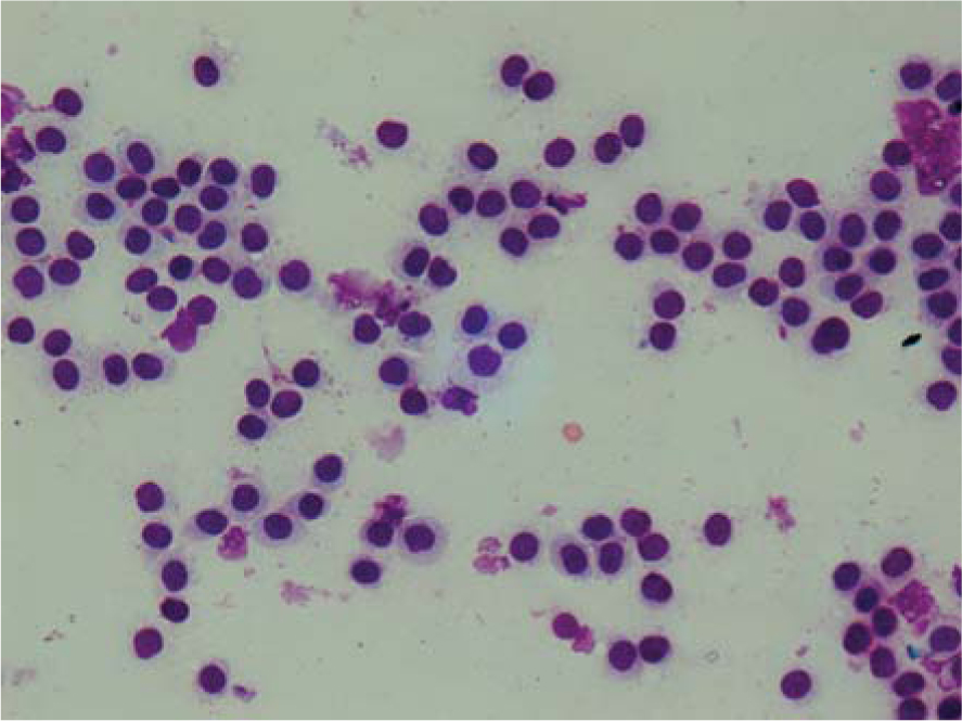
Figure 2
Cytological presentation of mast cell tumours (Tumour 2). Hemacolor, 400 x.
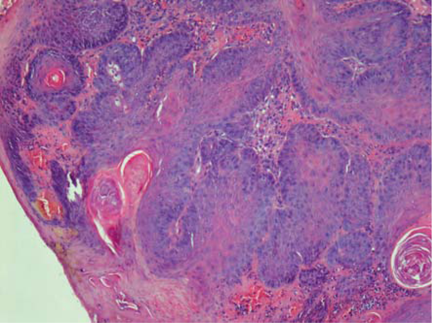
Figure 3
Histological presentation of squamous papilloma (Tumour 4). Hematoxylin and eosin stain, 100 x.
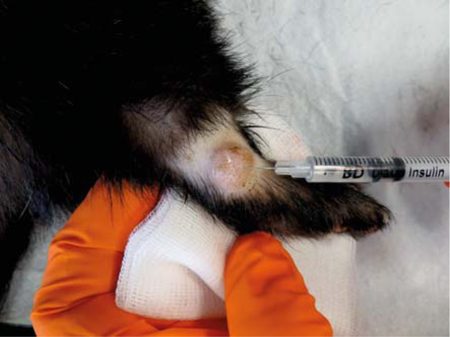
Figure 4
Intralessional injection of bleomycin into mast cell tumour (Tumour 2) located laterally on the right hock. Note the size of tumor and surgicaly chalenging anatomical location.
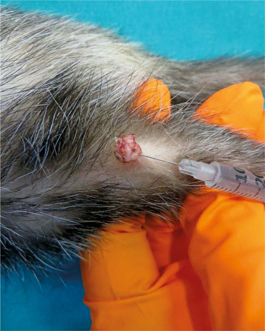
Figure 5
Intralessional injection of bleomycin into squamous papilloma (Tumour 4).
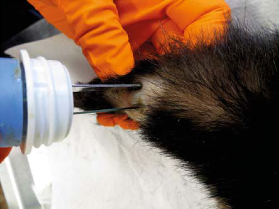
Figure 6
Electric pulses were delivered to the mast cell tumour (Tumour 2) using plate electordes.
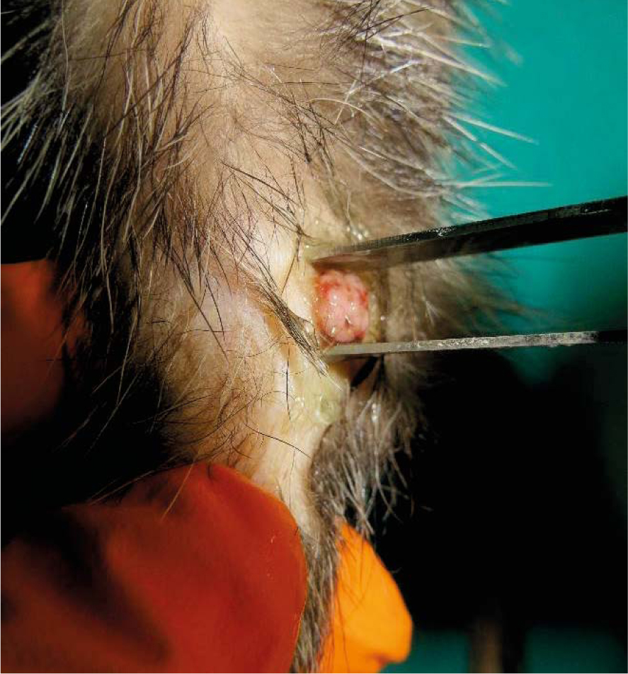
Figure 7
Electric pulses were delivered to the squamous papilloma (Tumour 4) using plate electordes.
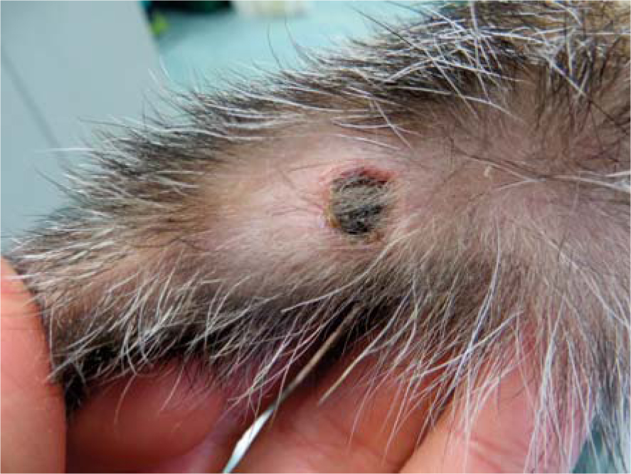
Figure 8
N ecrosis with superficial scab of squamous papilloma (Tumour 4) 8 days after electrochemotherapy.
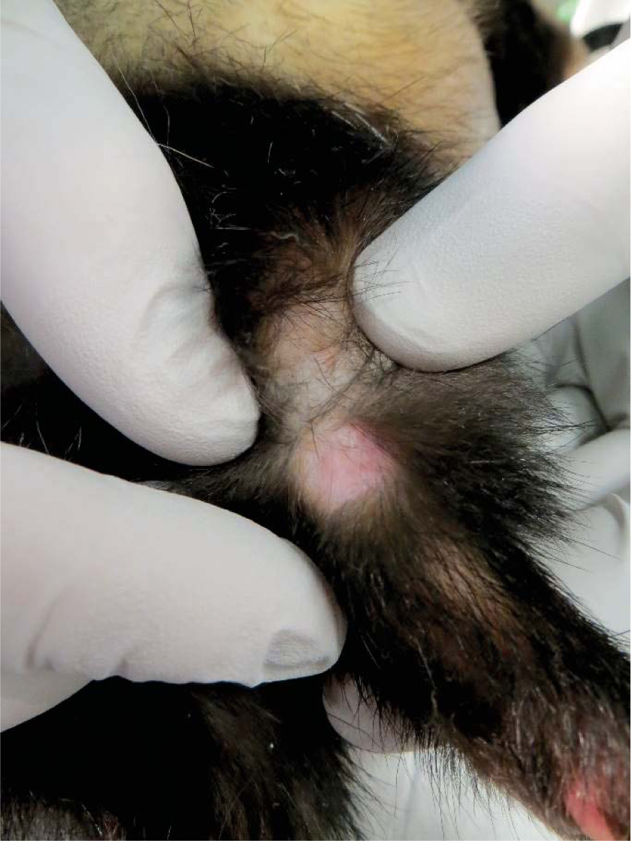
Figure 9
Complete response of mast cell tumour (Tumour 2) - 67 days after electrochemotherapy.
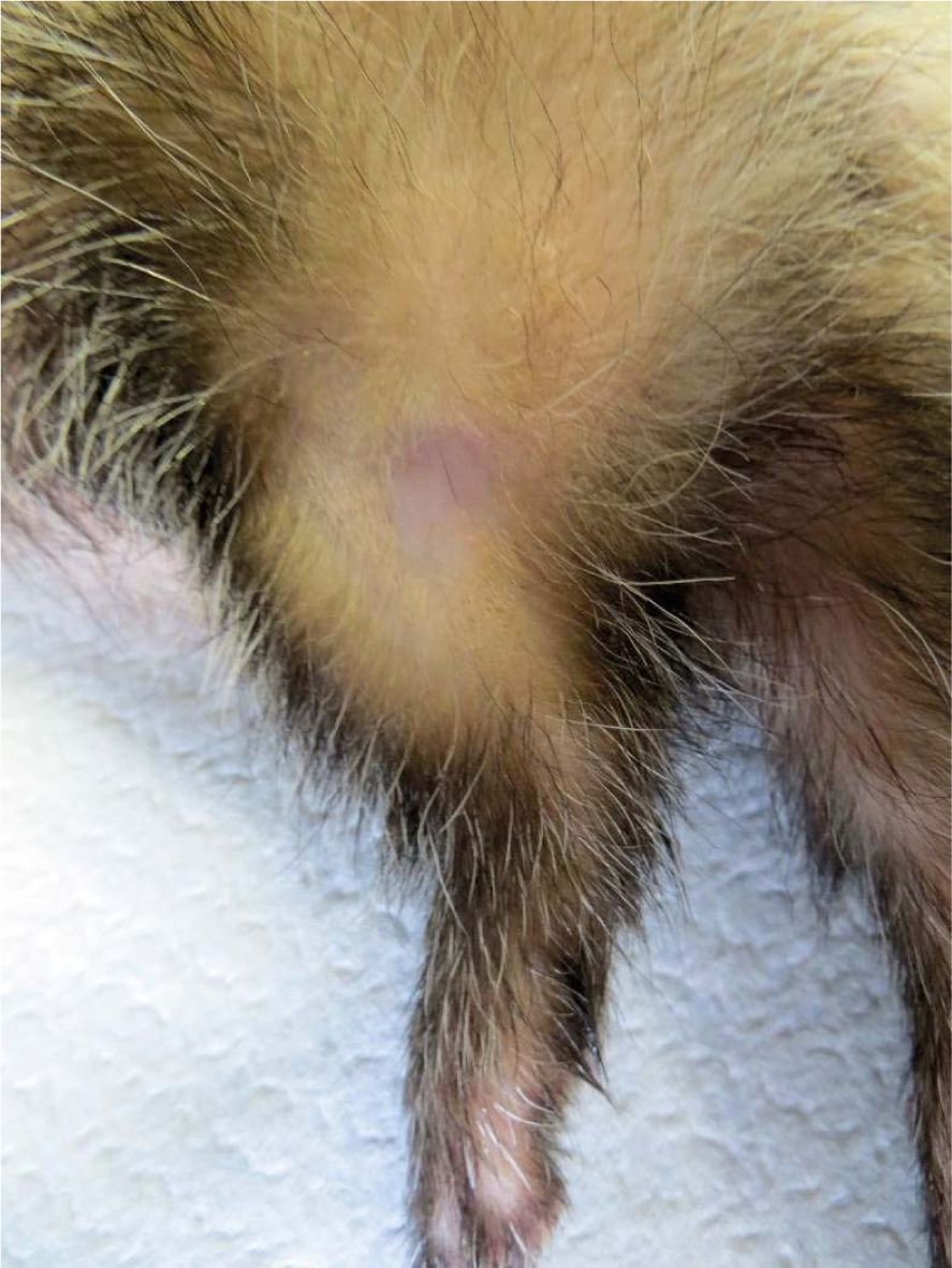
Figure 10
Complete response of squamous papilloma (Tumour 4) - 70 days after electrochemotherapy.