
Table 1
Patients’ demographic characteristics and radiological findings
| Mean age, years | 70.47±5.5 |
|---|---|
| Gender, no. | |
| Female | 54 |
| Male | 46 |
| Osteoarthritis, n | |
| Mild (Grade I and II) | 55 |
| Severe (Grade III and IV) | 45 |
| Chondromalacia patella, n | |
| Normal | 25 |
| Mild (Grade I and II) | 29 |
| Severe (Grade III and IV) | 46 |
| Medial meniscal tear, no | |
| Normal | 4 |
| Degeneration | 45 |
| Tear | 51 |
| Bone marrow edema, no | 45 |
| Others | |
| Enchondroma | 4 |
| Synovial chondromatosis | 4 |
| Osteochondroma | 1 |
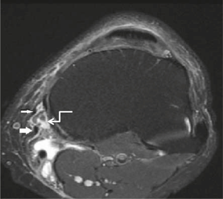
Figure 1
A 70 year-old patient presented with knee pain. Axial MR image shows sartorius (thin arrow) and gracilis tendon (thick arrow) in the patient with pes anserine bursitis (curved arrow).
Table 2
Locations of bursitis determined in the knee joint
| Bursae | No. |
|---|---|
| PA bursitis | 26 |
| MCL bursitis | 13 |
| SM-TCL bursitis | 12 |
| Iliotibial bursitis | 5 |
| LCL bursitis | 2 |
| Prepatellar bursitis | 4 |
| Infrapatellar bursitis | 4 |
| Suprapatellar bursitis | 3 |
| Baker’s cyst | 36 |
[i] LCL = lateral collateral ligament; MCL = medial collateral ligament; PA = Pes Anserine; SM-TCL = semimembranosus–tibial collateral ligament
Table 3
The relation between osteoarthritis, chondromalacia patella, meniscal tear and medial periarticular bursitis
| Medial compartment bursitis | |||
|---|---|---|---|
| Yes | No | p | |
| Osteoarthritis | |||
| Mild (n = 55) | 17 | 38 | 0.023 |
| Severe (n = 45) | 24 | 21 | |
| Chondromalacia Patella | |||
| Normal (n = 25) | 8 | 17 | |
| Mild (n = 29) | 14 | 15 | 0.479 |
| Severe (n = 46) | 19 | 27 | |
| Meniscal Tear | |||
| No (n = 49) | 15 | 34 | 0.038 |
| Yes (n = 51) | 26 | 25 | |
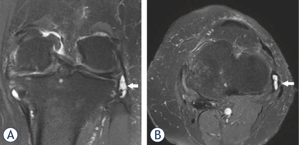
Figure 2
Coronal (A) and axial (B) proton-density fat-suppressed images of knee show LCL bursitis in a 70 years old patient.
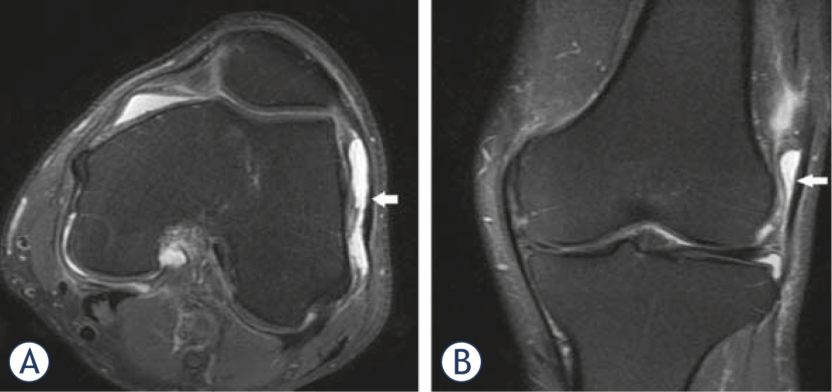
Figure 3
A 80 year-old patient presented with pain and tenderness in knee joint. Axial (A) and coronal (B) images demonstrate a fluid collection in the dilated iliotibial bursa.
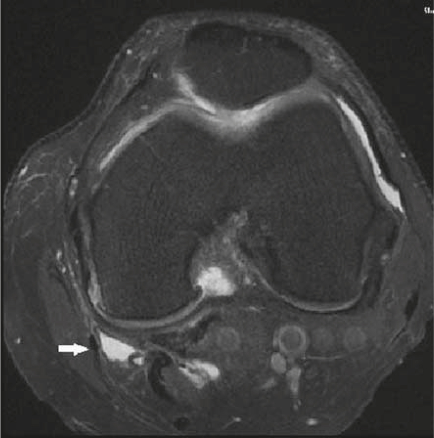
Figure 4
The axial fat saturated proton density weighted image demonstrates a fluid collection in the SM-TCL bursa.