
Figure 1
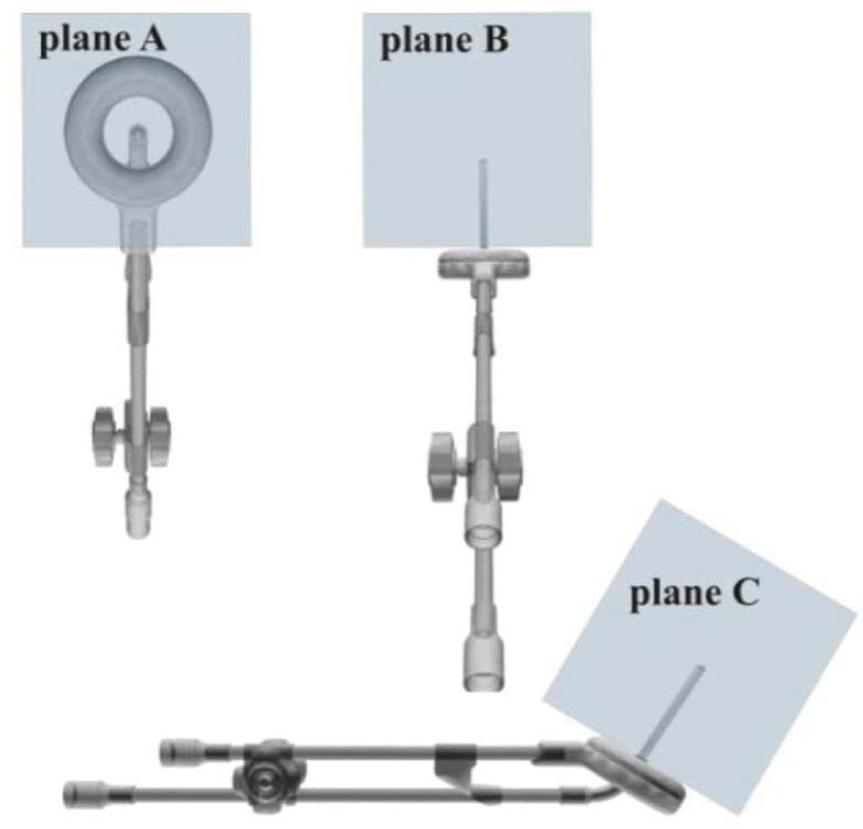
Figure 2
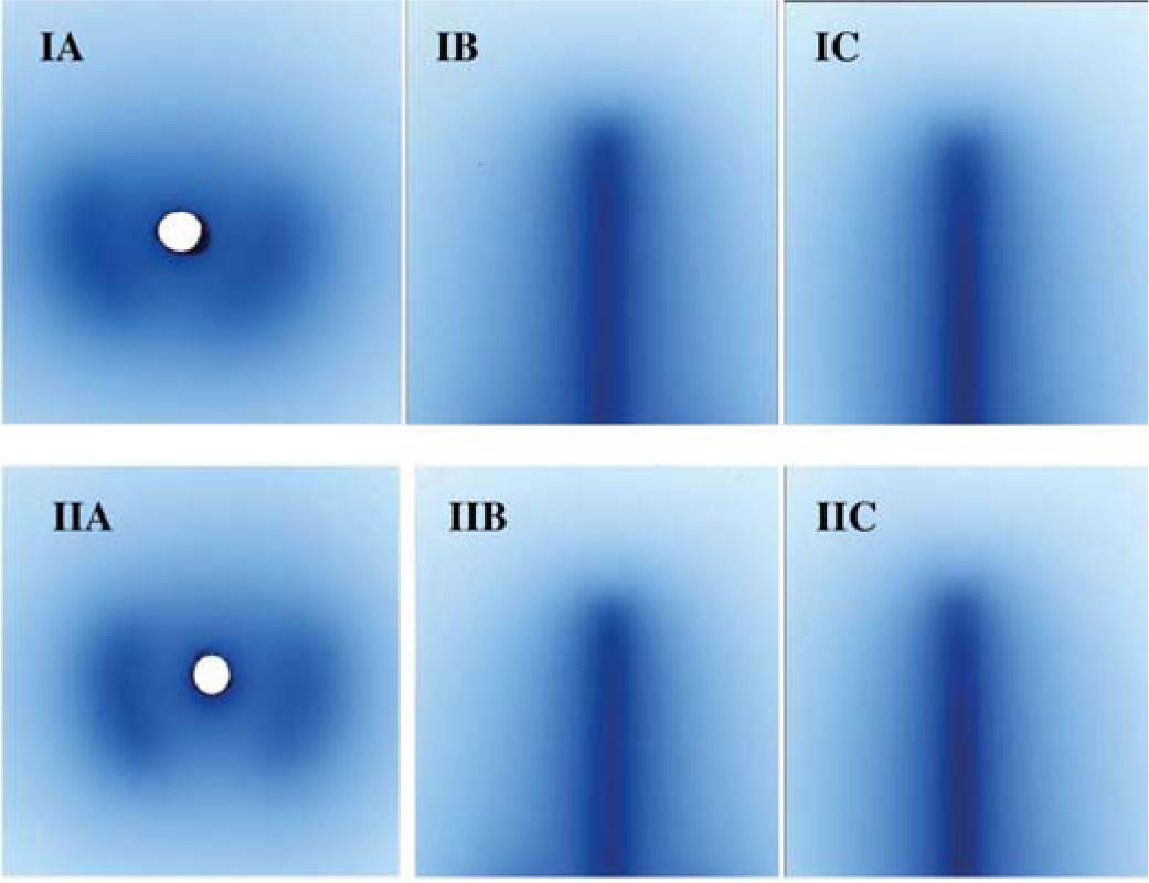
Figure 3
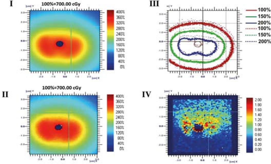
Figure 4
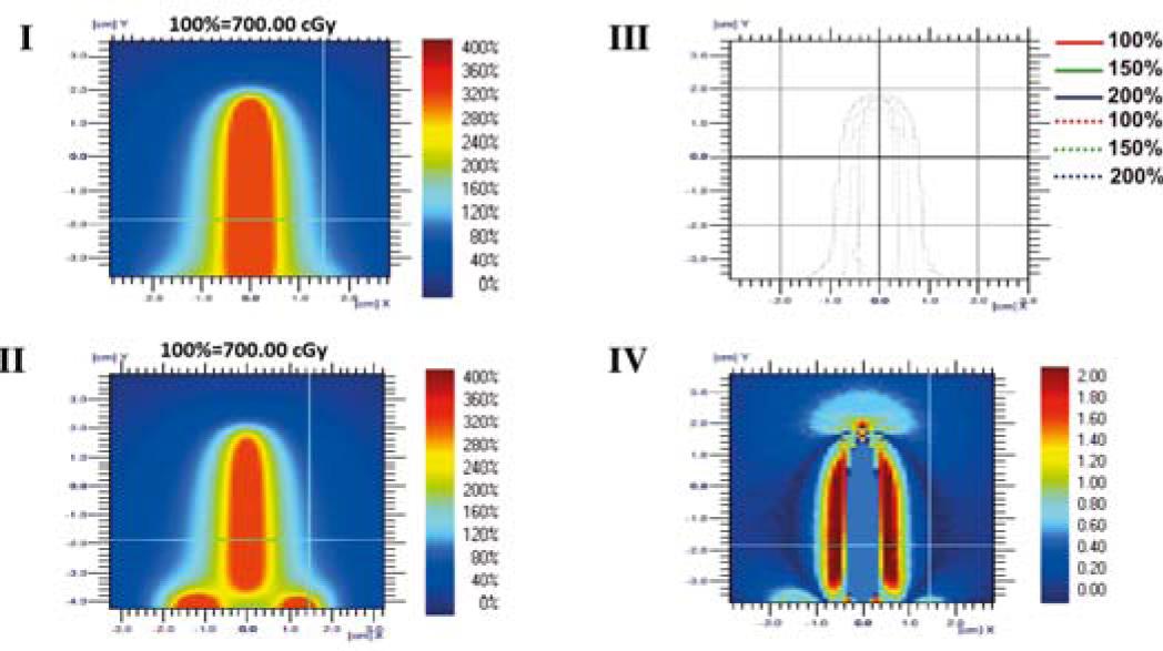
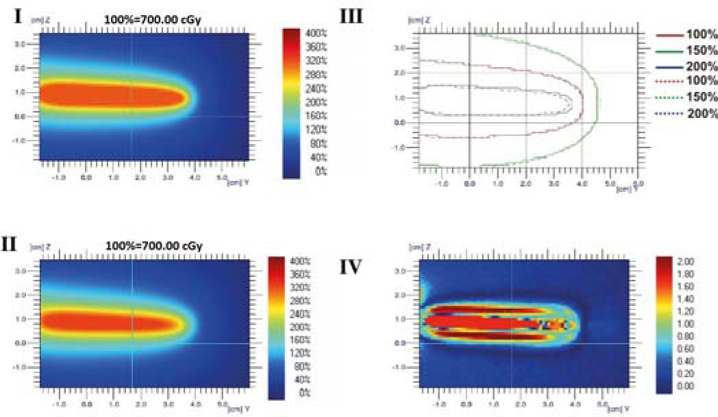
Figure 5
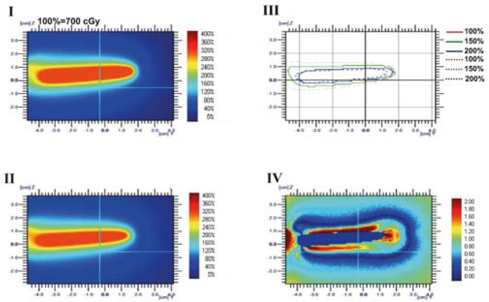
Figure 6
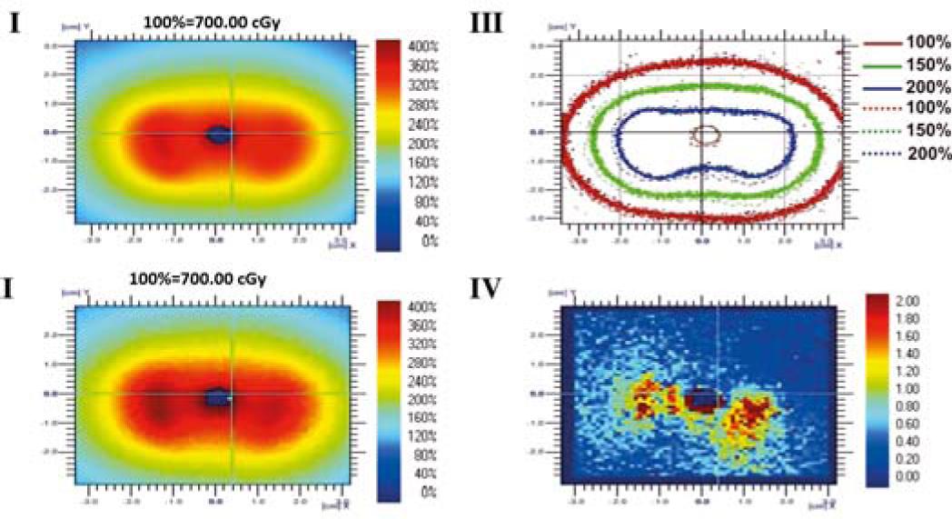
Figure 7
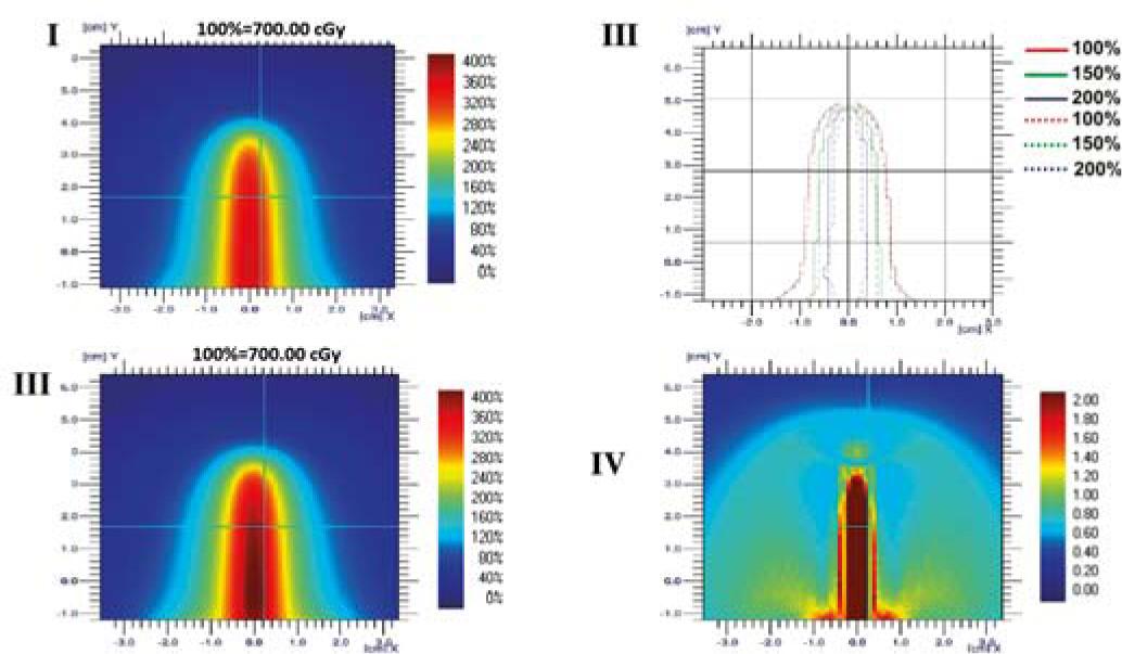
Figure 8
Results of Gamma analysis for the 26 mm ring and 30 mm ring applicator
| Ring, 26 mm | Gamma average | Gamma max | Gamma (0.00 – 1.00) | |
|---|---|---|---|---|
| plane A | 0.22 | 1.42 | 99.04% | |
| plane B | 0.10 | 1.29 | 99.31% | |
| plane C | 0.44 | 1.75 | 98.88% | |
| Ring, 30 mm | Gamma average | Gamma max | Gamma (0.00 – 1.00) | |
| plane A | 0.25 | 1.96 | 98.11% | |
| plane B | 0.27 | 2.00 | 97.94% | |
| plane C | 0.10 | 1.22 | 99.54% | |