
Table 1
Patients by age, size and position of an aneurysm, complications, and follow-up
| No/Age (years)/Gender | Clinical presentation | Location | Type | Size (mm)/Neck (mm) | Dilatation of the stent with the PTA balloon | Late clinical complications | Type of control to confirm closure/Status (month) | OKM Scale |
|---|---|---|---|---|---|---|---|---|
| 1/57/Female | Mass effect (vision loss) | Parophthalmic | S | 12/5 | – | – | CTA/Complete (3) | C3 |
| 2/72/Female | Incidental (headache) | Parophthalmic | S | 7/5 | – | – | CTA/Complete (14) | A2 |
| 3/55/Female | Recurrent (coil) incidental (headache) | Parophthalmic | S | 5/3 | – | – | CTA/Complete (5) | B3 |
| 4/47/Female | Incidental (headache) | Petrous ICA | S | 4/4 | PTA | – | CTA/Complete (6) | B3 |
| 5/62/Female | Incidental (headache) | ICA – PCOM | S | 6/6 | – | – | CTA/Complete (2) | D |
| 6/57/Female | Mass effect (vision loss) | Carotidcavernous | S | 7/4 | – | – | CTA/Complete (1) | D |
| 7/56/Female | Mass effect (diplopia) | Parophthalmic | S | 12/7 | PTA | – | CTA/Complete (3) | C2 |
| 8/48/Female | Mass effect (diplopia) | Parophthalmic | S | 17/7 | – | Stent shortening | MRA/Complete (15) | A3 |
| 9/22/Female | Mass effect (diplopia) | Parophthalmic | F | 20/10 | – | – | MRA/Complete (6) | B2 |
| 10/52/Female | Mass effect (diplopia) | ICA – PCOM | S | 23/9 | – | – | MRA/Complete (6) | B3 |
| 11/52/Female | Mass effect (diplopia) | Parophthalmic | S | 20/3 | – | – | DSA/Complete (3) | C2 |
| 12/55/Female | Mass effect (diplopia) | Parophthalmic | S | 22/5 | PTA | – | MRA/Complete (6) | C1 |
| 13/47/Female | Incidental (headache) | ICA – PCOM | S | 4/3 | – | – | DSA/Complete (3) | C3 |
| 14/54/Male | Recurrent aneurysm (clip) (headache) | ICA – cisternal | F | 16/14 | – | – | MRA/Complete (6) | B2 |
| 15/28/Male | Incidental (headache) | M1 | S | 6/5 | – | – | MRA/Complete (6) | B3 |
CTA = computed tomography angiography; DSA = digital subtraction angiography; ICA = internal carotid artery; MRA = magnetic resonance angiography; OKM = O‘Kelly-Marotta; PCOM = posterior communicating artery, PTA = percutaneous transluminal angioplasty
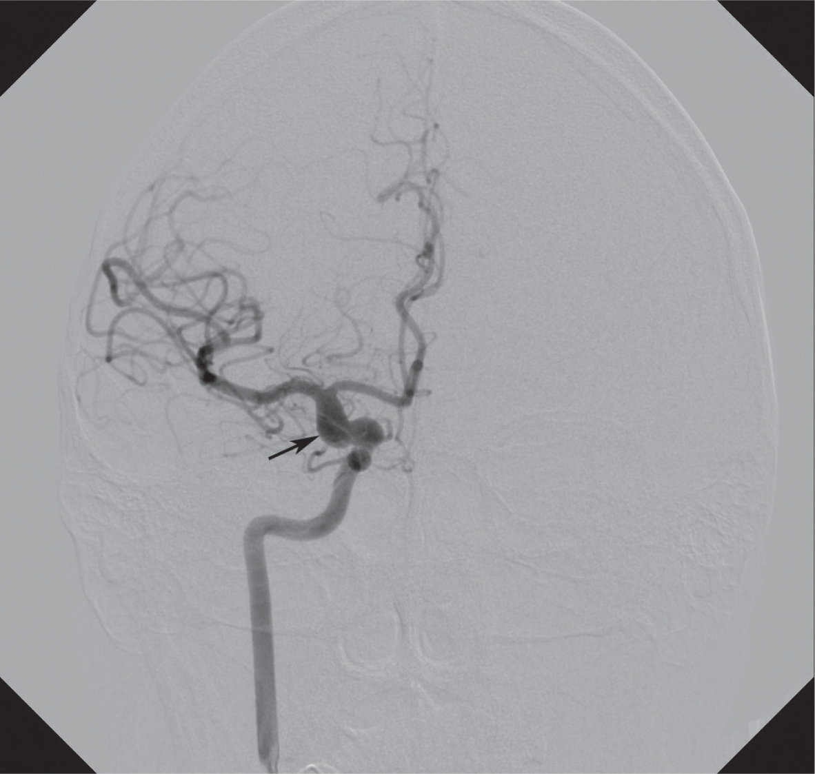
A fusiform aneurism of the internal carotid artery is indicated by the arrow.
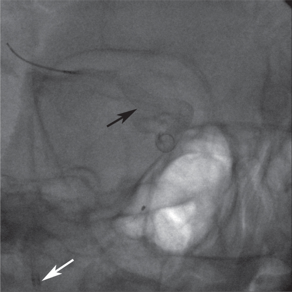
The position of the flow diverter stent on the native radiogram is indicated by the black arrow. The white arrow indicates the tip of the guiding catheter.
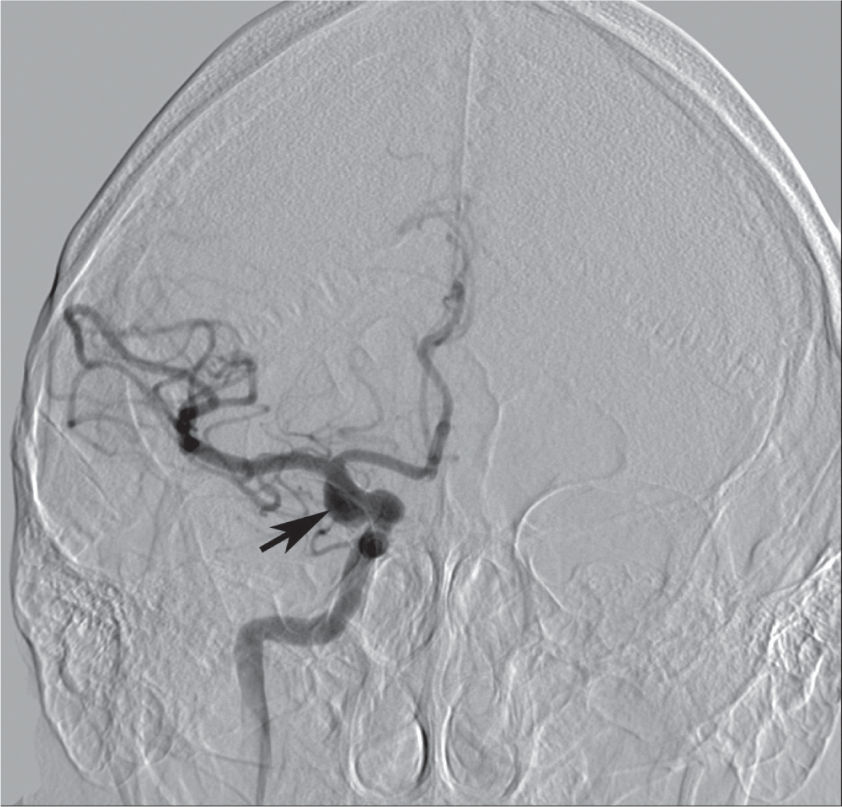
Control DSA direct after the positioning of the flow diverter stent shows some residual filling of the aneurism (arrow). The stent fits the vessel wall perfectly.
Contril DSA after 12 months shows complete thrombosis of the aneurism (arrow) and good patency of the distal arteries.
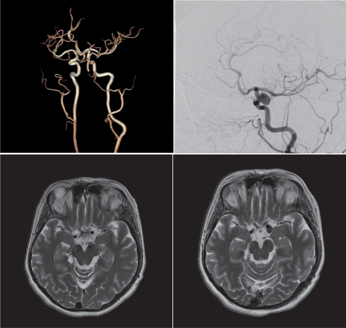
Control MR after 12 months shows complete thrombosis of the aneurism (arrow) and aneurysm collapse following flow diverter treatment.