
Table 1.
Patients’ characteristics
| Patient 1 | Patient 2 | Patient 3 | Patient 4 | Patient 5 | |
|---|---|---|---|---|---|
| Gender | Male | Male | Female | Female | Female |
| Birth year | 1958 | 1935 | 1937 | 1953 | 1941 |
| TNM* | T3aNOMO | T2aNOMO | T4bN1aMO | T3aN1aMO | T4bN1aMO |
| Patological stage* | Stage IIa | Stage Ib | Stage IIIb | Stage IIIa | Stage IIIb |
| Breslow | 4 mm | 1.5 mm | 10 mm | 2.8 mm | 9.5 mm |
| Ulceration | / | / | 10 mm | / | 2.5 mm |
| Localisation of primary tumor | Right lower leg | Left foot | Right foot | Right lower leg | Back |
| Localisation of metastases treated with ECT | Right lower leg | Left foot (dorsum) | Left lower leg | Right lower leg | Breast, left side |
* according to AJCC TNM Cancer Staging Manual 7th edition (2010)6; ECT = electrochemotherapy
* according to AJCC TNM Cancer Staging Manual 7th edition (2010)6; ECT = electrochemotherapy
Table 2.
Treatment regime
| Patient 1 | Patient 2 | Patient 3 | Patient 4 | Patient 5 | |
|---|---|---|---|---|---|
| INF-a dose | 6 million IU 3 × weekly (s.c.) | 3 million IU 3 × weekly (s.c.) | 35 milion IU 20 × (i.v.) in 8 weeks/15 million IU 3× weekly (s.c.) for 13 weeks/ 10 milion IU 3 × weekly (s.c.) for 13 weeks* | 30 milion IU 20 × (i.v.) in 4 weeks followed by 15 million IU 3× weekly (s.c.) for 35 weeks followd by 10 milion IU 3 × weekly (s.c.) for 12 weeks | 30 milion IU 9 × (i.v.) in 2 weeks followed by 20 milion IU 11× (i.v.) in 3 weeks followed by 15 million IU 3× weekly (s.c.) for 13 weeks** |
| INF-α treatment period | 2 years 9 months | 7 months | 8 months | 12 months | 4 months |
| Last date of INF-α treatment | March 2004 | August 1998 | July 2012 | January 2012 | June 2011 |
| Disease free interval | 4 months | 5 years 6 months | 6 months | 3 months | 2 months |
| Surgically excised metastases++ | Yes | Yes | No | Yes | Yes |
| Interval between INF-α and ECT treatment | 4 years 8 months | 12 years 11 months | 7 months | 6 months | 7 months |
| ECT treatment | November 2008 | July 2011 | February 2013 | July 2013 | January 2012 |
| ECT drug | Cisplatin | Cisplatin | Bleomycin | Bleomycin | Bleomycin |
| Number of lesions | 2 | 1 | 80 | 23 | 5 |
| Size of the nodules | 1.0 × 1.5 cm 1.5 × 1.5 cm | 3 × 3 cm | 0.3–1 cm | 0.1–0.8 cm | 0.7–1.5 cm |
| Effect after 4 weeks | CR | CR | > 85% CR | All CR | 100% PR |
| LRD*** or date of death (D) | April 2010 (LRD) | December 2014 (LRD) | April 2014 (LRD) | December 2013 (D) | March 2013 (D) |
CR = complete response; ECT = electrochemotherapy; i.v. = intravenous; PR = partial response; s.c. = subcutaneously; s.c = subcutaneous;
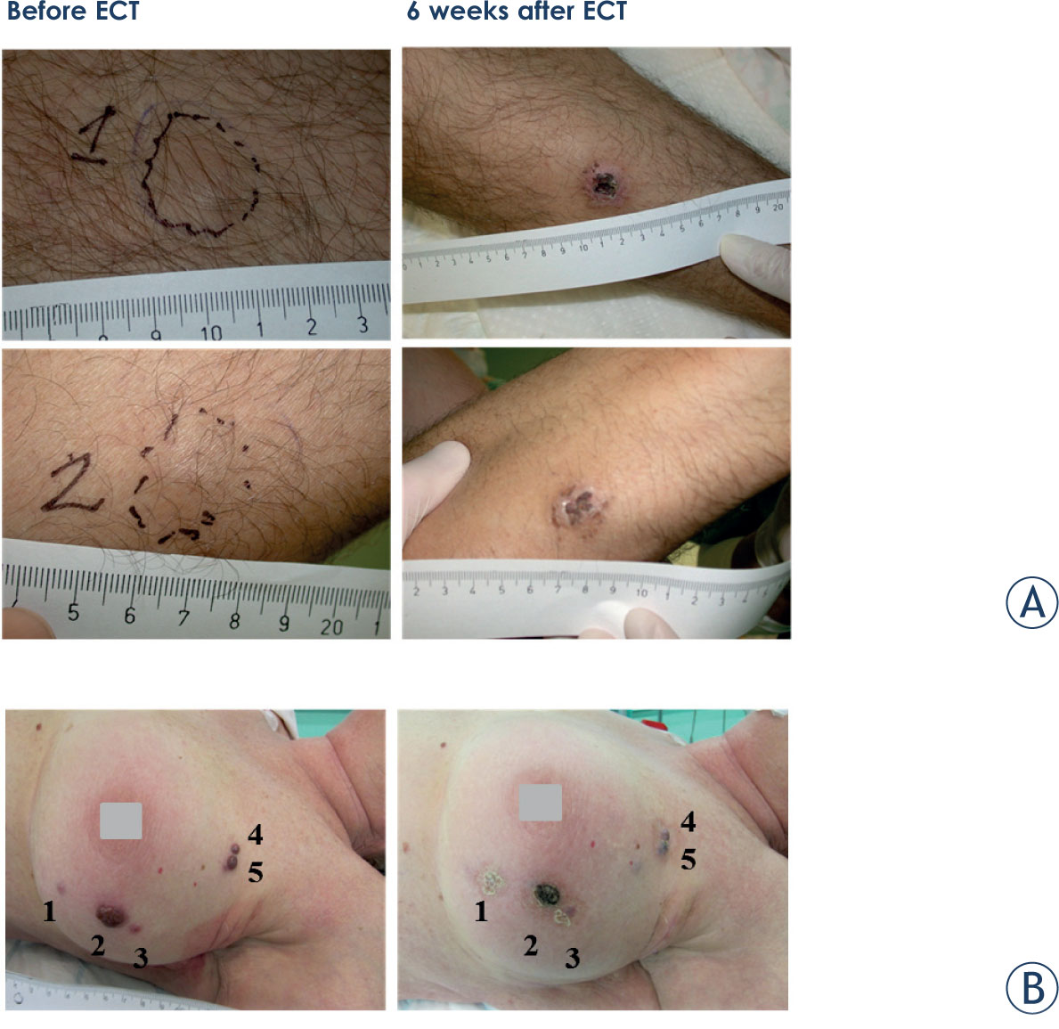
Figure 1.
Melanoma lesions in patient 1 (A) and patient 5 (B) before and 6 weeks after electrochemotherapy (ECT). Both metastases in patient 1 responded completely, while in patient 5 although the crust is seen on the images there was nodule under it and response was evaluated as partial response.