
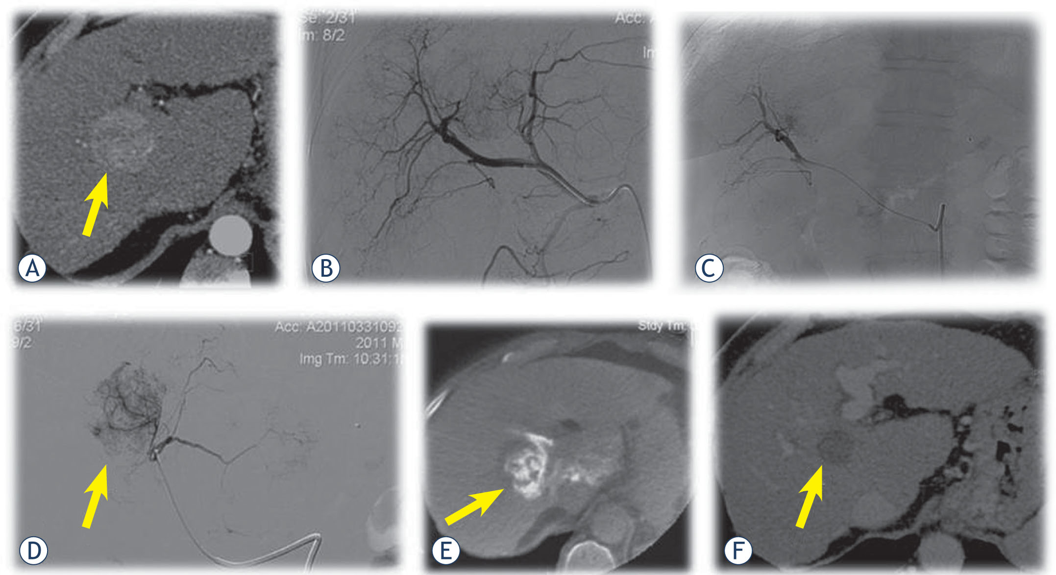
A 56-year-old female with HCC. (A) Contrast-enhanced CT shows tumor (arrow) between right and left liver lobe. (B) Initial angiography shows tumor in the liver (arrow). (C) Superselective contrast injection throught microcatheter into segmental branches for eight liver segment confirmes the tumor. (D) Superselective contrast injection throught microcatheter into segmental branches for first liver segment also confirmes the tumor (arrow). (E) CBCT after contrast injection throught microcatheter into segmental branches for first liver segment confirmes that the target lesion (arrow) is dominantely suplied from this artery and superselective DEBDOX TACE was performed from both artery. (F) Two months after the DEBDOX TACE, control CT shows complete devascularization of the target lesion (arrow) (complete response).
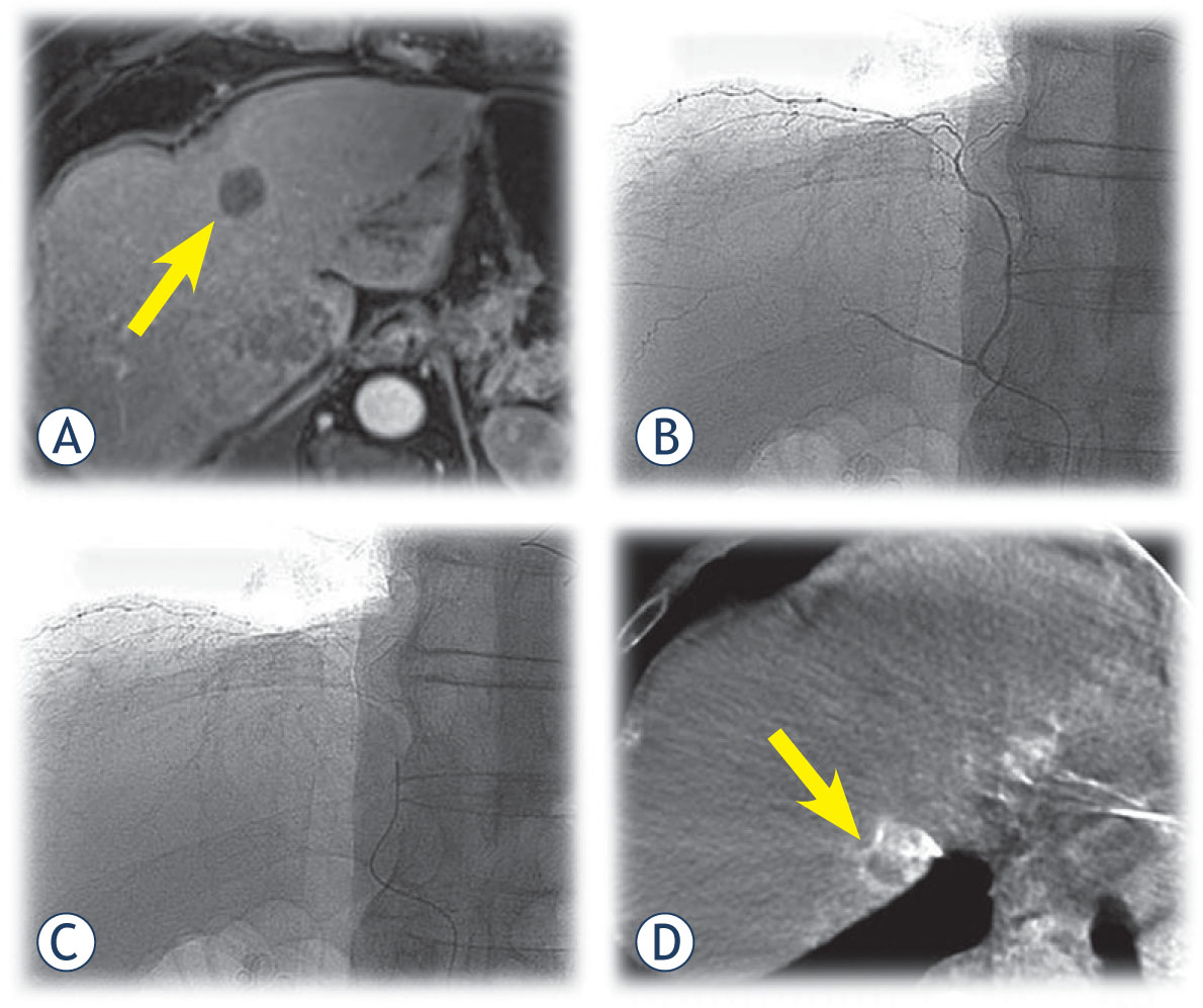
A 64-year-old male with HCC. (A) Control MR after chemoembolization shows complete response of the target lesion (arrow) in fourth segment and partial response of the target lesion in eight segment. (B) Superselective contrast injection throught microcatheter into the righr phrenic artery. (C) Position of microcatheter prior CBCT. (D) CBCT shows that this artery suplied the target tumor in seventh segment. Superselective DEBDOX TACE was performed.
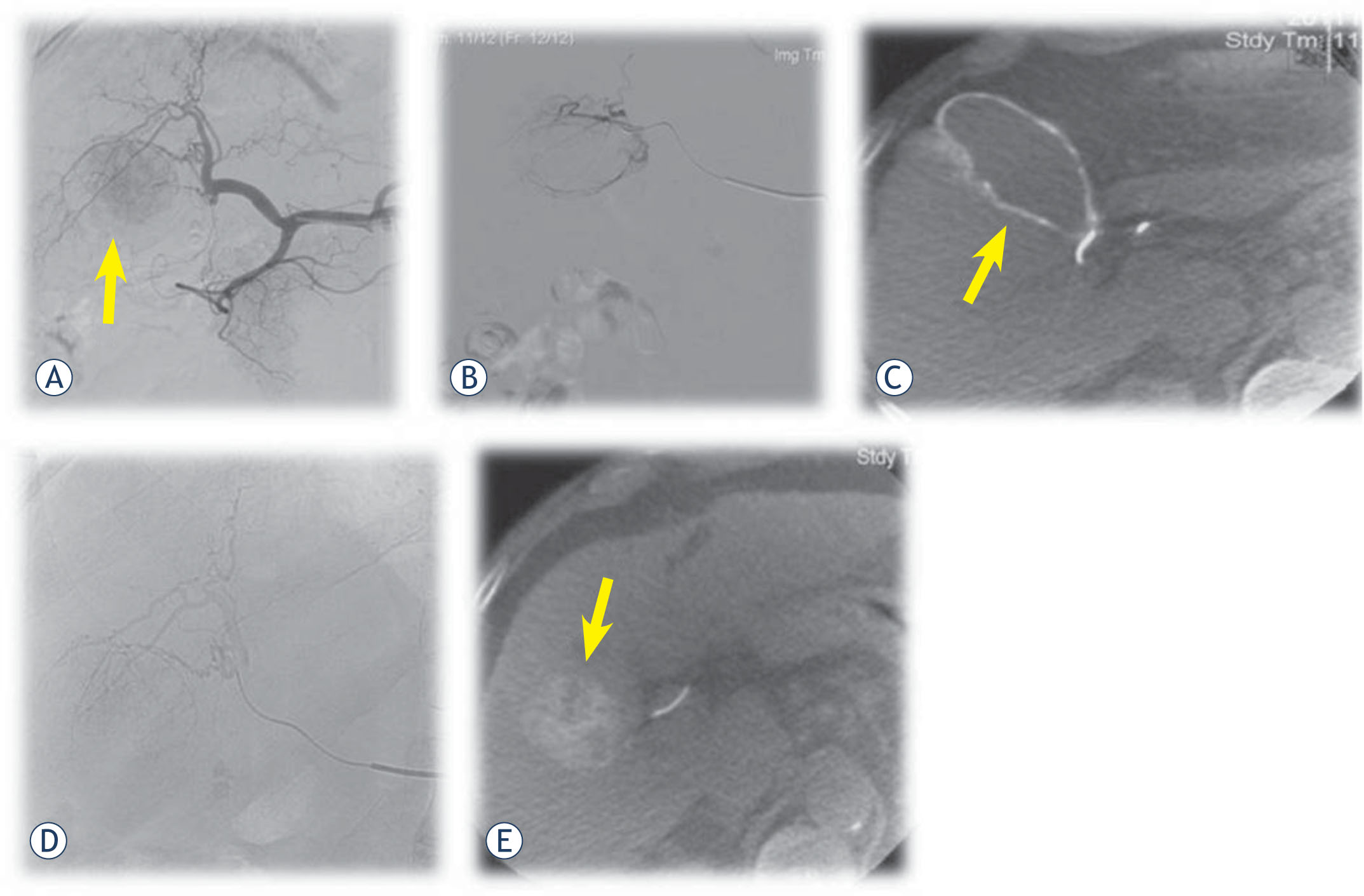
A 66-year-old male with HCC. (A) Initial angiography shows tumor in right liver lobe (arrow). (B) Superselective contrast injection throught microcatheter into the subsegmental branches. (C) CBCT shows that this artery actually suplied the gallblader (arrow) and not the target tumor. (D) Superselective catheterisation of the tumor feeding artery with microcathter. (E) CBCT after contrast injection throught microcatheter confirmes the target lesion (arrow). Superselective DEBDOX TACE was performed.
Table 1
Baseline demographic, clinical, laboratory and tumor staging characteristics of patients
| Characteristic | Value |
|---|---|
| Age, [years] | 67.5 ± 7.8 |
| Gender (M/F), n [%] | 32/3 [91.4/8.6] |
| Cirrhosis (yes/no), n [%] | 30/5 [85.7 /14.3] |
| Etiology of cirrhosis, n [%]: | |
| Ethanol | 13 [43.3] |
| HBV | 4 [13.3] |
| HCV | 4 [13.3] |
| other | 9 [29.9] |
| Albumin [g/l] | 38.4 ± 4.6 |
| INR | 1.2 ± 0.2 |
| Total bilirubin [μmol/l] | 25.6 ± 17.4 |
| Child-Pugh score (points) | 6.0 ± 0.7 |
| Child-Pugh score (classes), n [%]: | |
| A | 22 [73.3] |
| B | 8 [26.7] |
| Creatinine [μmol/l] | 81.7 ± 23.4 |
| ASAT [μkat/l] | 1. 1 ± 0.7 |
| ALAT [μkat/l] | 0.8 ± 0.6 |
| γGT [μkat/l] | 2.3 ± 1.6 |
| αFP [kIE/l] | 152.5 ± 310.2 |
| Portal vein thrombosis (yes/no), n [%] | 6/29 [17.1/82.9] |
| Bilobar disease, n [%] | 11 [31.4] |
| Unilobar disease, n [%:] | 24 [68.6] |
| right lobe, n [%] | 20 [83.3] |
| Overall number of nodules, n | 97 |
| Number of nodules per pt, n | 2.8 ± 2.2 |
| Maximum diameter of HCC nodule per pt. [cm] | 4.7 ± 1.9 |
HBV = hepatitis B virus, HCV = hepatitis C virus; INR = international normalised ratio; ASAT = aspartate aminotransferase; ALAT = alanine aminotransferase; γGT = gamma glutamyl transpeptidase; αFP = alpha-feto protein; pt = patient. Quantitative variables expressed as mean ± standard deviation.
Table 2
Number and type of minor complications after DEBDOX TACE.
| Type of complication | No. of complications |
|---|---|
| Post-embolization syndrome** | 23 |
| Rise in blood pressure | 4 |
| Gastric erosions or ulcers | 2 |
| Chest pain | 2 |
| Hematoma at puncture site | 1 |