
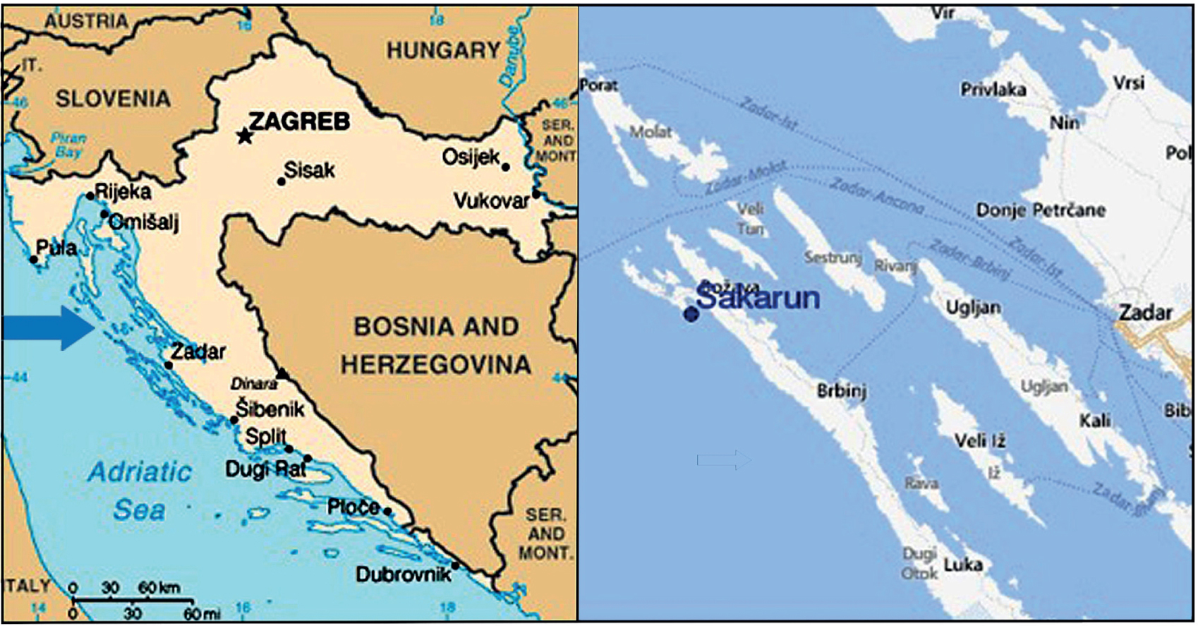
Figure 1
Sampling site in Croatia, Dugi Otok island at Saharun Bay, the Adriatic Sea (Google maps)
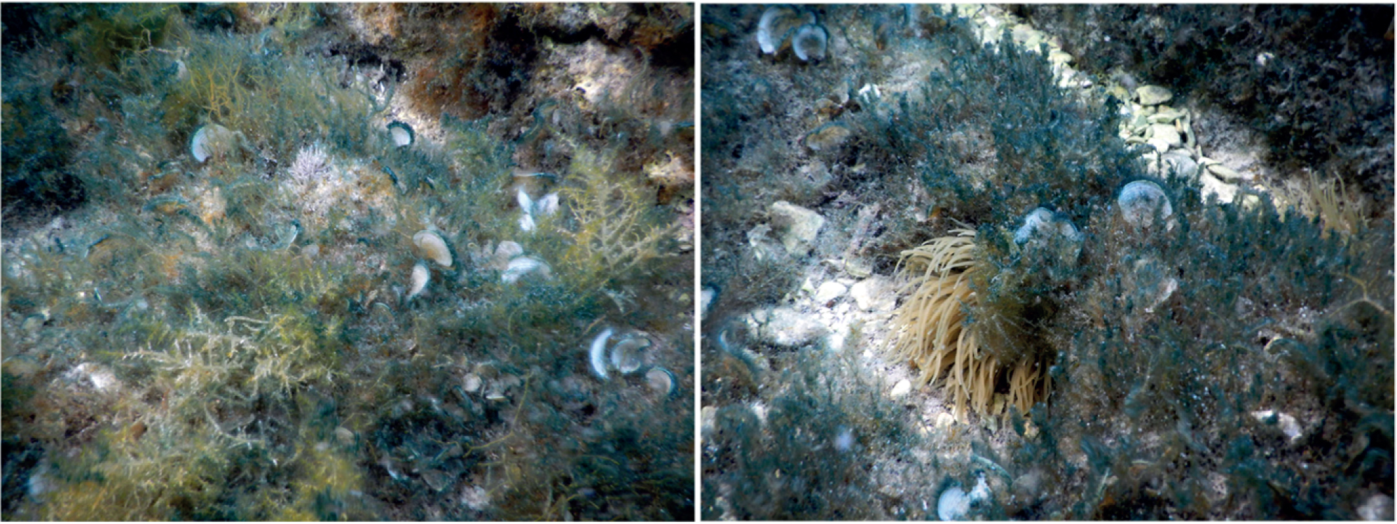
Figure 2
Field population of Haslea sp. Typical bluish color of diatoms attached to Padina sp. is clearly visible
Table 1
Morphological features of Haslea sp. (this study) compared with those of H. ostrearia, H. provincialis, H. karadagensis, H. crucigera and H. pseudostrearia obtained from the literature
| Haslea sp. | Haslea ostrearia | Haslea provincialis | Haslea karadagensis | Haslea crucigera | Haslea pseudostrearia | |
|---|---|---|---|---|---|---|
| Length (μm) | 60.1 | 71.8 | 65.8 | 52.5 | 55.5 | 95–97 |
| Width (μm) | 7.6 | 7.3 | 7.4 | 8.0 | 8.8 | 11–121 |
| Color of cell apices | blue | blue | blue | blue-grey | no | no |
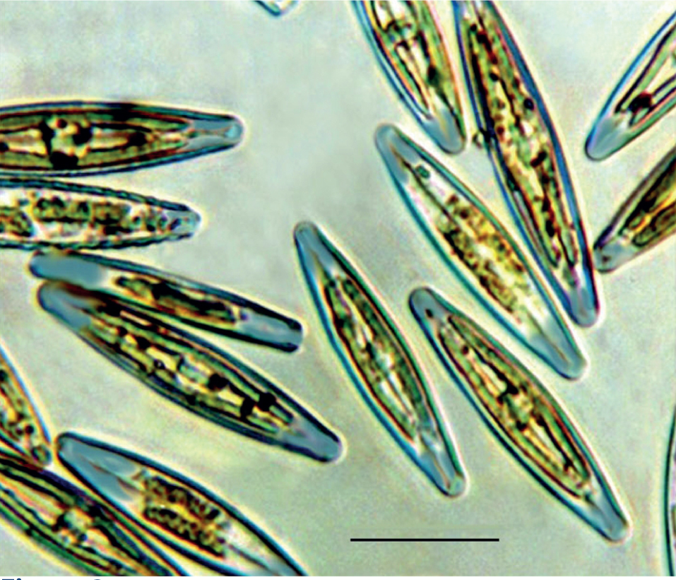
Figure 3
Light microscopy of Haslea sp., with parietal chloroplasts and apices filled with blue pigment. Scale bar = 30 μm
Table 2
18S rRNA gene fragments sequence similarities (%) among Haslea strains
| Haslea sp. (this study) | Haslea ostrearia | Hasle pseudostrearia | Haslea crucigera | Haslea spicula BA28 | |
|---|---|---|---|---|---|
| H. ostrearia | 97.16 | ||||
| H. pseudostrearia | 94.34 | 98.06 | |||
| H. crucigera | 94.33 | 97.99 | 97.75 | ||
| H. spicula BA28 | 94.09 | 98.35 | 98.06 | 98.87 | |
| H. sp. RG-2015a | 99.70 | 96.96 | 93.64 | 93.62 | 93.31 |