
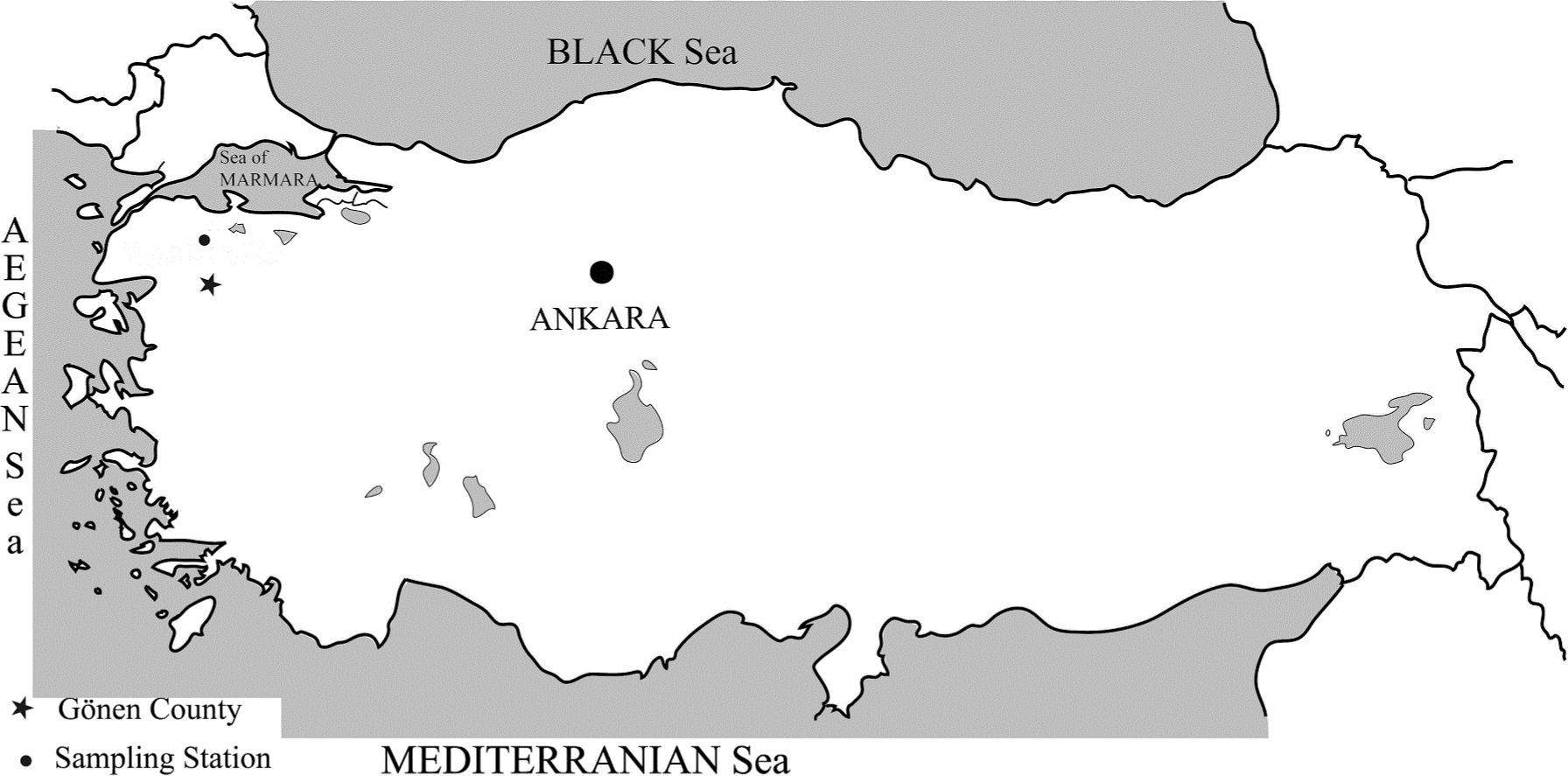
Figure 1
Location of the sampling station in Turkey
Table 1
Physicochemical factors, coordinates and altitude of habitats with their species
| Habitat | T (°C) | S (PSU) | EC (µS) | DO (mg l-1) | %S (%) | pH | Coordinate | Alt (m) | Species (Numbers) |
|---|---|---|---|---|---|---|---|---|---|
| Rice field | 21.6 | 0.3 | 657 | 5.9 | 67 | 7.75 | 40°11′08″N | 66 | Hv (34), Sb (3), Bp (5), Hr (715) |
| Spring | 18.2 | 0.2 | 370 | 8.6 | 94 | 8.11 | 27°31′15″E | Hv (38), Sb (3), Hr (58), Hi (2), Hs (8) |
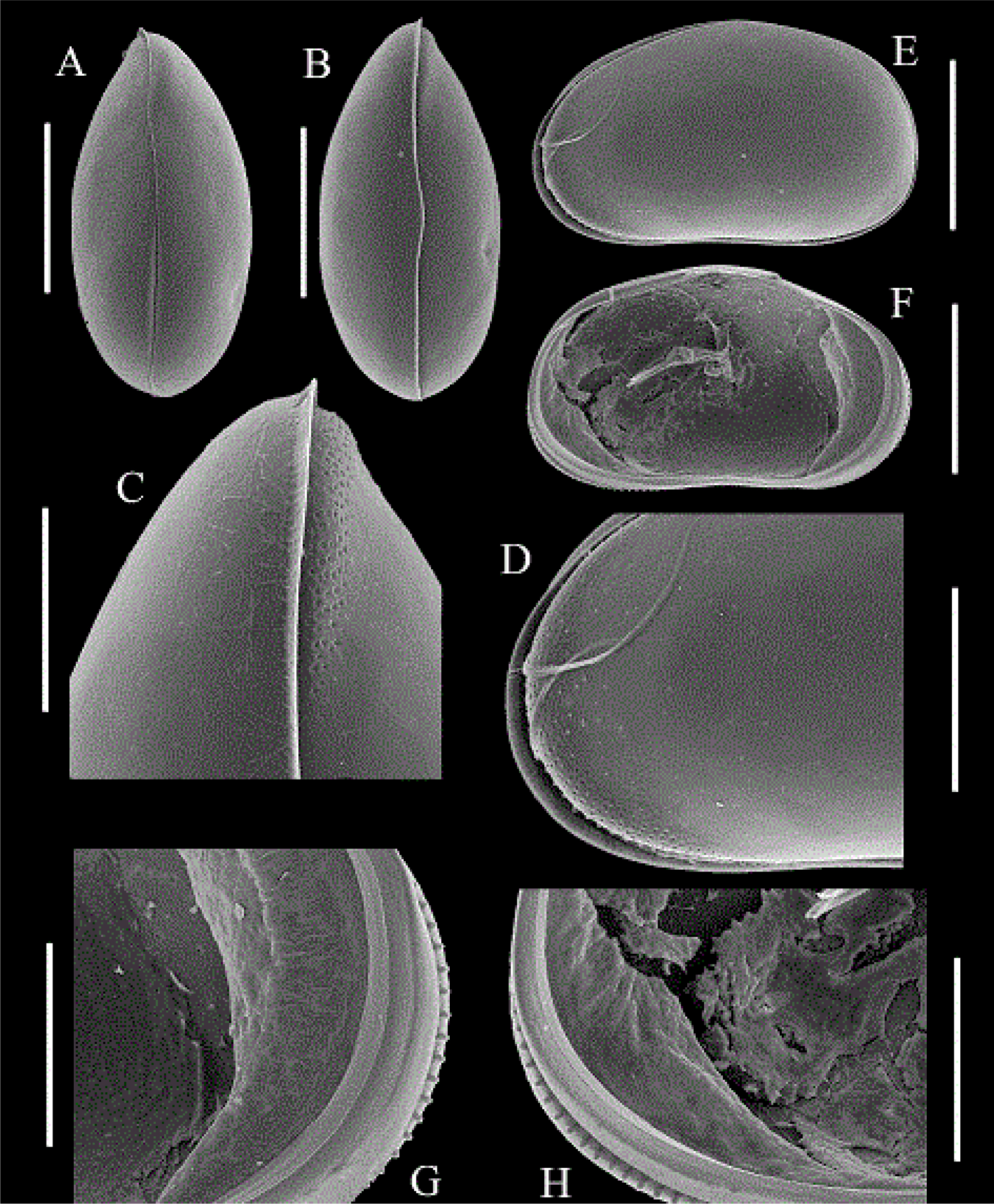
Figure 2
Hemicypris vulgaris Okubo, 1990 (Female). A: Dorsal view of carapace; B: Ventral view of carapace; C: Anterior part of carapace (ventral view); D-E: external view of the left valve (anterior – whole); F-H: Internal view of the left valve (whole – anterior – posterior) and part of the left valve; External view of the left valve. Scales (μm): A, B, E, F = 500; C, D, G, H = 200
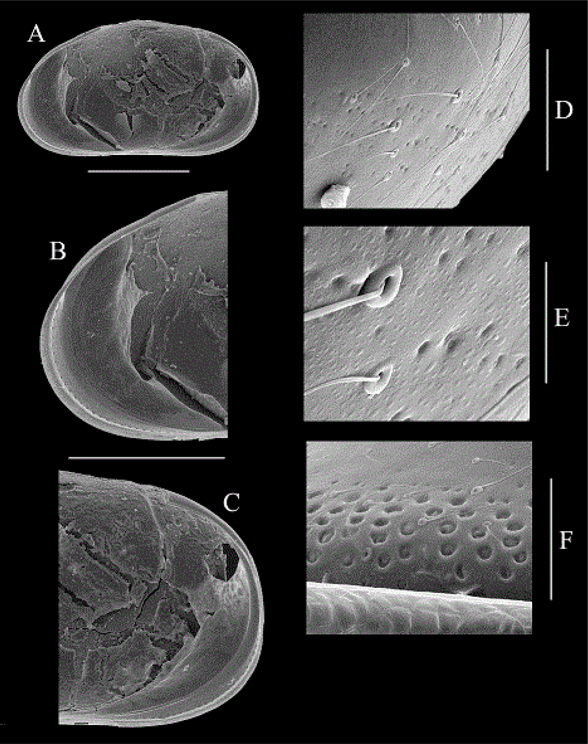
Figure 3
Hemicypris vulgaris Okubo, 1990 (Female). A-C: Interior view of the right valve (whole – anterior – posterior); D-E: Surface of carapace and pore canals; F: Pitted area of the anterior end of the left valve. Scales (μm): A = 500; B-C = 400; D-F = 40; E = 10
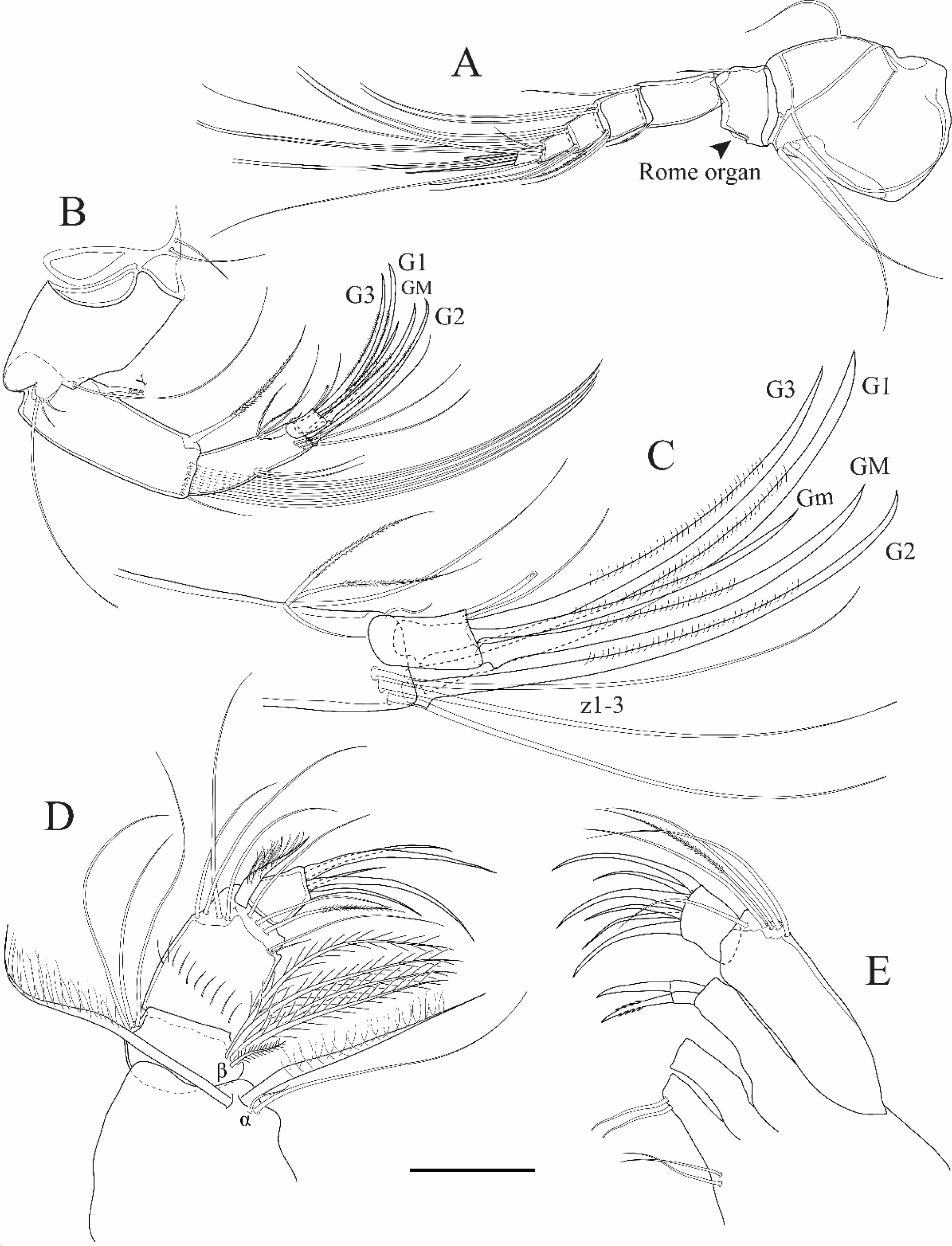
Figure 4
Hemicypris vulgaris Okubo, 1990 (Female). A: Antennula; B-C: Antenna (whole – last segments); D: Mandibula; E: Maxillula (other setae on endites omitted here). Scales (μm): A = 80; B = 110; C = 30; D-E = 140
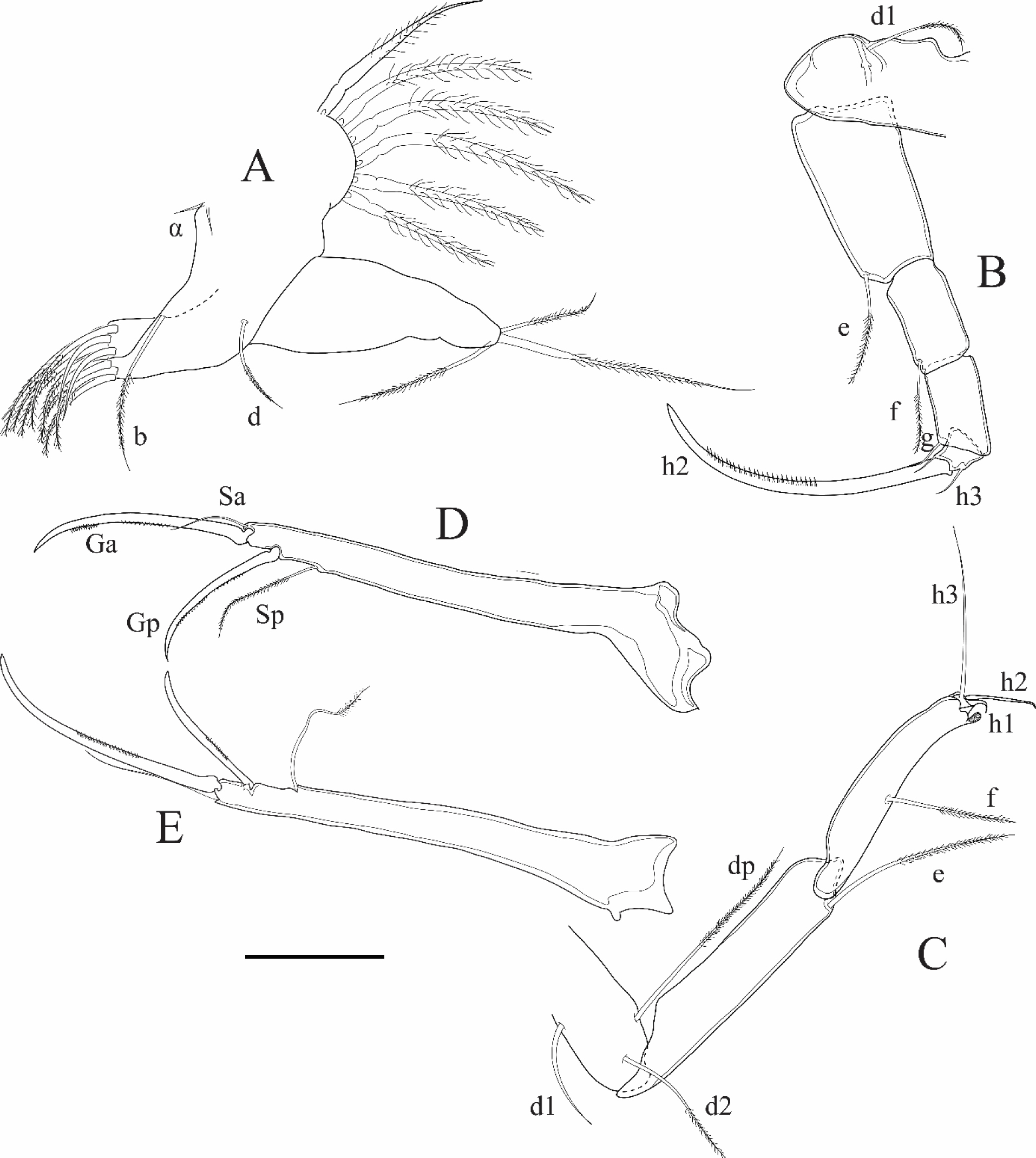
Figure 5
Hemicypris vulgaris Okubo, 1990 (Female). A: First thoracopod; B: Walking leg; C: Cleaning leg; C: Cleaning leg; D: Uropodal ramus; E: Hemicypris ovata Sars, 1903 (Femaile, Lectotype) uropodal ramus. Scales (µm): A-C = 140; D-E = 110
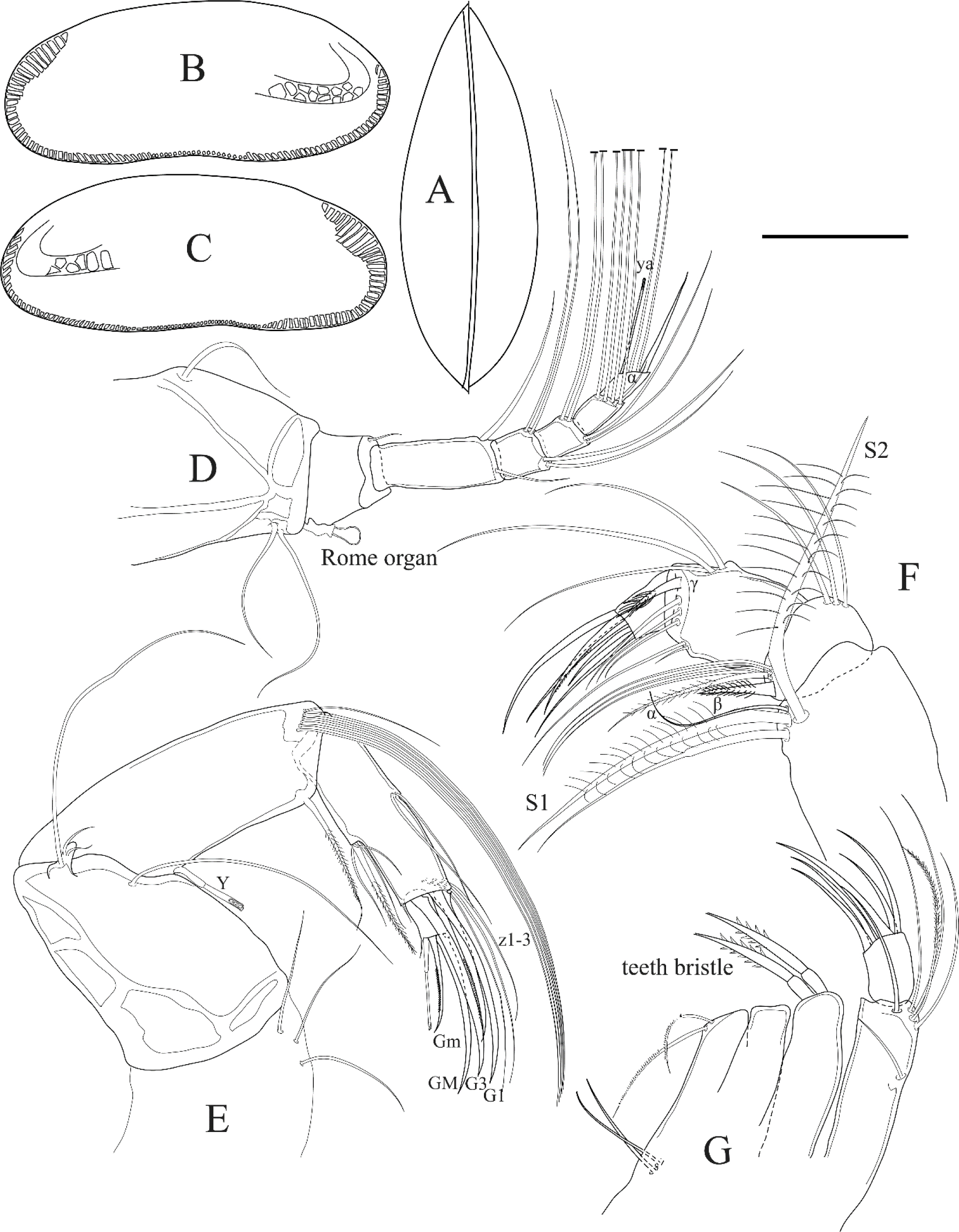
Figure 6
Stenocypris bolieki Ferguson, 1962 (Female). A: Left valve (outer side); B: Right valve (outer side); C: Dorsal view; D: Antennula; E: Antenna; F: Mandibula. G: Maxillula (other setae are omitted here). Scales (μm): A-C = 680; others: 68
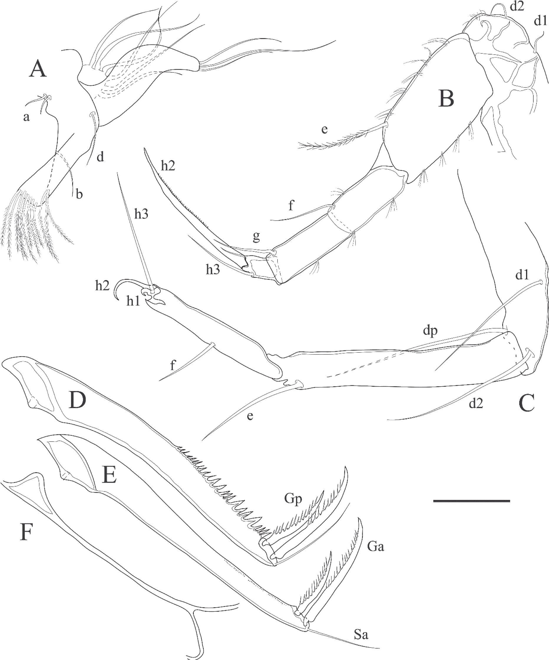
Figure 7
Stenocypris bolieki Ferguson, 1962 (Female). A: First thoracopod; B: Walking leg; C: Cleaning leg; D-E: Uropodal rami (left-right); F: Uropodal attachment. Scale: 68 μm
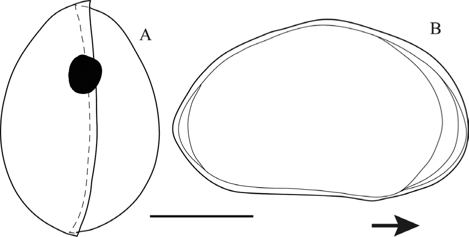
Figure 8
Bradleystrandesia parva (Hartmann, 1964) (Female). A: Dorsal view of carapace; B: Internal view of the left valve. Scales (μm): A = 300; B = 250
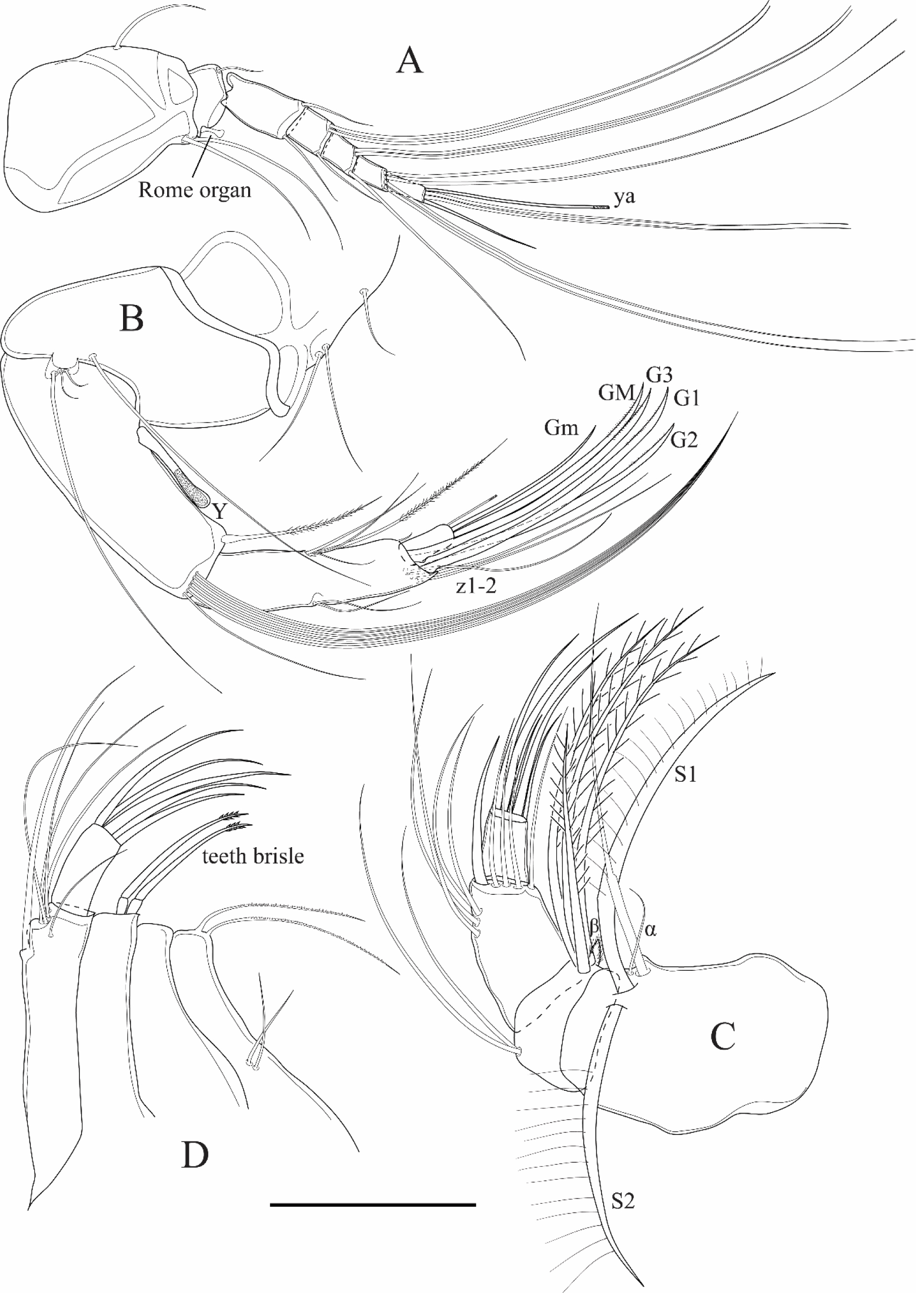
Figure 9
Bradleystrandesia parva (Hartmann, 1964) (Female). A: Antennula; B: Antenna C: Mandibula; D: Maxillula (other setae on endites are omitted here). Scales (μm): A-B = 52; C-D = 70
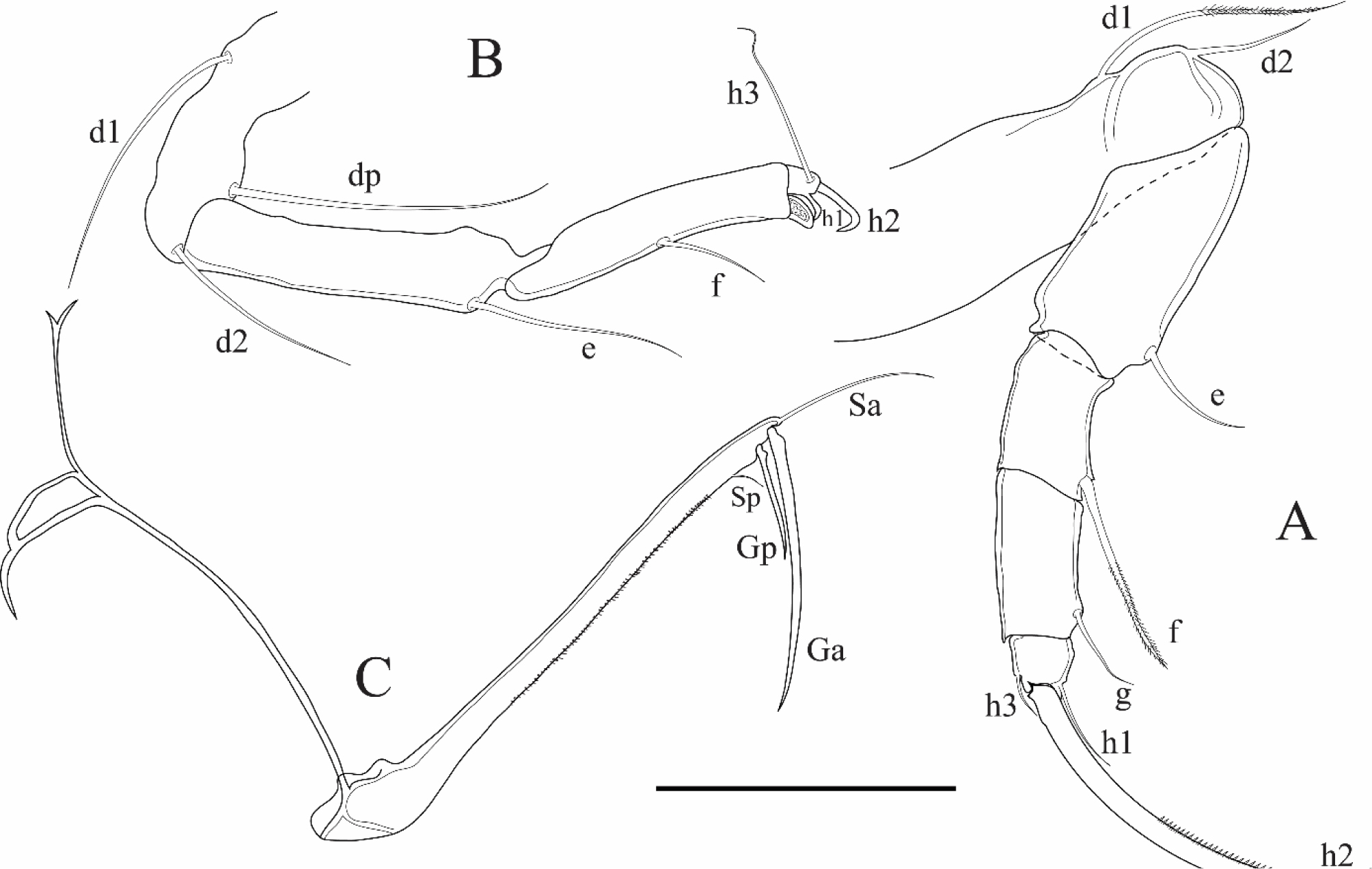
Figure 10
Bradleystrandesia parva (Hartmann, 1964) (Female). A: Walking leg; B: Cleaning leg; C: Uropodal ramus. Scales (μm): A = 52; B-C = 70