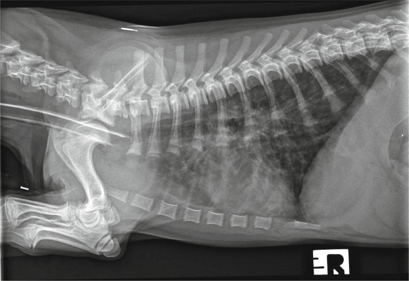
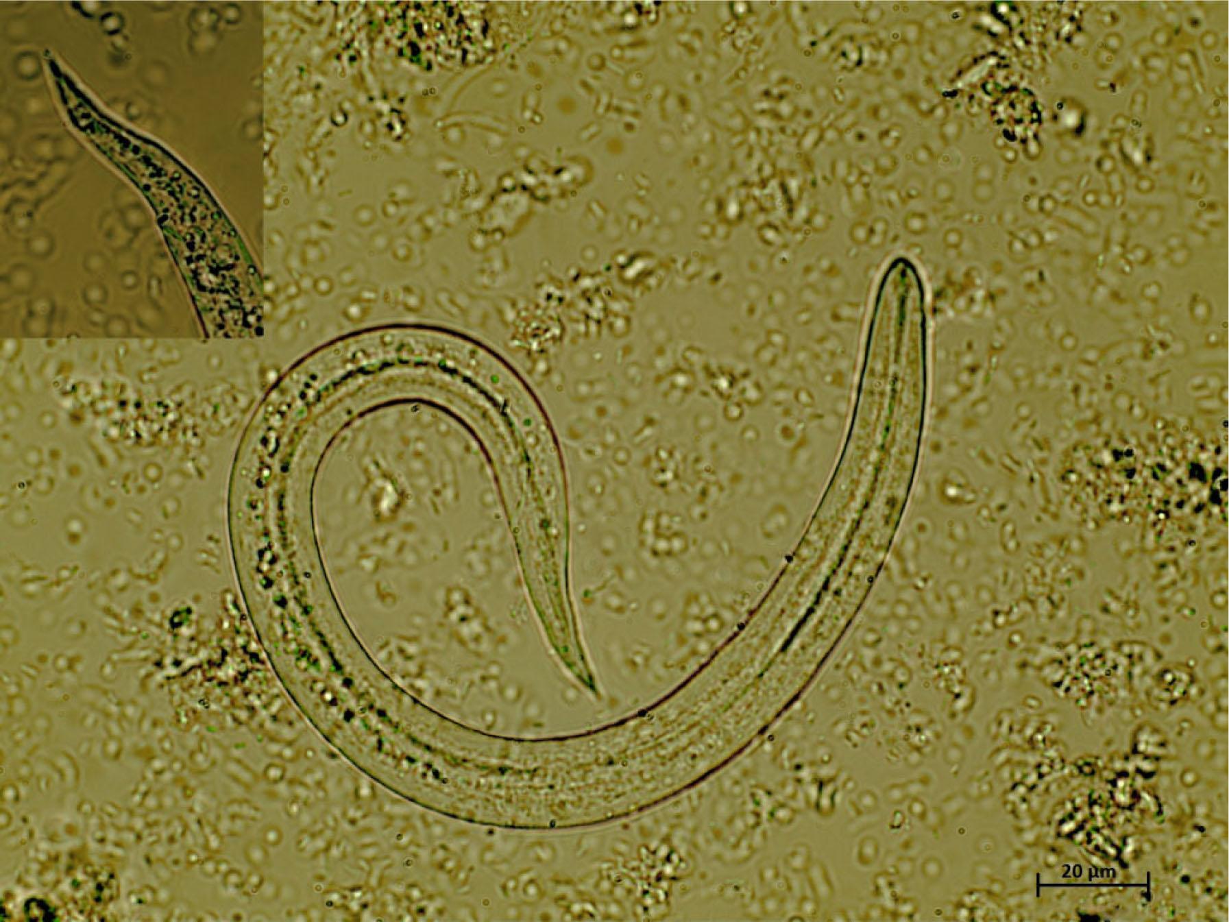
Have a personal or library account? Click to login

Crenosoma vulpis infection in a four-month old puppy

Open Access
|Jul 2016

Figures & Tables

Fig. 1

Right lateral thoracic radiograph of the dog showing thickening of the bronchial walls into the periphery of the lung fields and a diffuse increase in pulmonary opacity throughout the lungs

Fig. 2

Crenosoma vulpis first stage larvae detected at the Baermann technique, 40x magnification. Note the filiform C-shaped larvae with conical tail, consistent with C. vulpis
DOI: https://doi.org/10.1515/helmin-2016-0027 | Journal eISSN: 1336-9083 | Journal ISSN: 0440-6605
Language: English
Page range: 276 - 280
Submitted on: May 2, 2016
Accepted on: Jun 8, 2016
Published on: Jul 22, 2016
Published by: Slovak Academy of Sciences, Mathematical Institute
In partnership with: Paradigm Publishing Services
Publication frequency: 4 times per year

© 2016 B. Matos, V. Colella, A. M. Alho, D. Otranto, R. Doyle, L. Madeira de Carvalho, published by Slovak Academy of Sciences, Mathematical Institute
This work is licensed under the Creative Commons Attribution-NonCommercial-NoDerivatives 3.0 License.