
Table 1
Morphometric data, in millimeters, of Amphiorchis solus (Simha & Chattopadhyaya, 1970) Platt, 2002 (Digenea, Spirorchiidae) from Chelonia mydas Testudines, Cheloniidae)
| Amphiorchis solus (Simha & Chattopadyaya, 1970) | |||
|---|---|---|---|
| Variables | Simha & Chattopadyaya (1970) | Wernecket al. (2011) | Presente report |
| Host | C. mydas | C. mydas | C. mydas |
| Locality | Camp Mandapam Gulf of Manar, Índia | Barra Nova Beach, State of Ceará, Brazil | Mangue Seco, state of Bahia, Brazil |
| Site of infection | Heart | Heart | Heart |
| Number of parasites | 1 | 1 | 1 |
| Body length (mm) | 3.96 | 2.86 | 2.85 |
| Body width anterior part* | 0.81 | 0.55 | 0.54 |
| Body width posterior part** | - | - | 0.44 |
| Oral sucker length | 0.17 | 0.16 | 0.11 |
| Oral sucker width | 0.16 | 0.14 | 0.09 |
| Acetabulum length | 0.25 | 0.32 | 0.29 |
| Acetabulum width | - | 0.23 | 0.29 |
| Esophagus length | 1.17 | 0.97 | 1.01 |
| Esophagus width | - | - | 0.079 |
| Anterior testis length | 0.58 | 0.31 | 0.35 |
| Anterior testis width | 0.51 | 0.31 | 0.25 |
| Posterior testis length | 0.26 | 0.20 | 0.13 |
| Posterior testis width | 0.27 | 0.17 | 0.25 |
| External seminal vesicle length | 0.23 | 0.18 | 0.11 |
| External seminal vesicle width | - | 0.091 | 0.083 |
| Cirrus pouch length | 0.22 | 0.26 | 0.16 |
| Cirrus pouch width | - | - | 0.066 |
| Ovary length | 0.32 | 0.14 | 0.22 |
| Ovary width | 0.30 | 0.13 | 0.15 |
| Distance from: | |||
| Genital pore to posterior end | - | - | 0.45 |
| Intestinal caeca bifurcation to anterior end | - | - | 1.1 |
| Intestinal cecal end to posterior end | - | - | 0.12 |
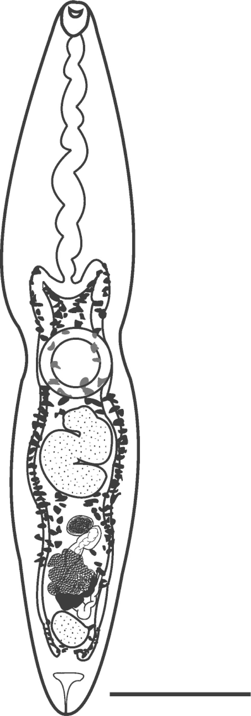
Fig. 1
Amphiorchis solus (Simha & Chattopadhyaya, 1970) Platt, 2002 (Digenea: Spirorchiidae) found in Chelonia mydas Linnaeus 1758 (Testudines, Cheloniidae) from Brazil. (scale bar = 500 μm)
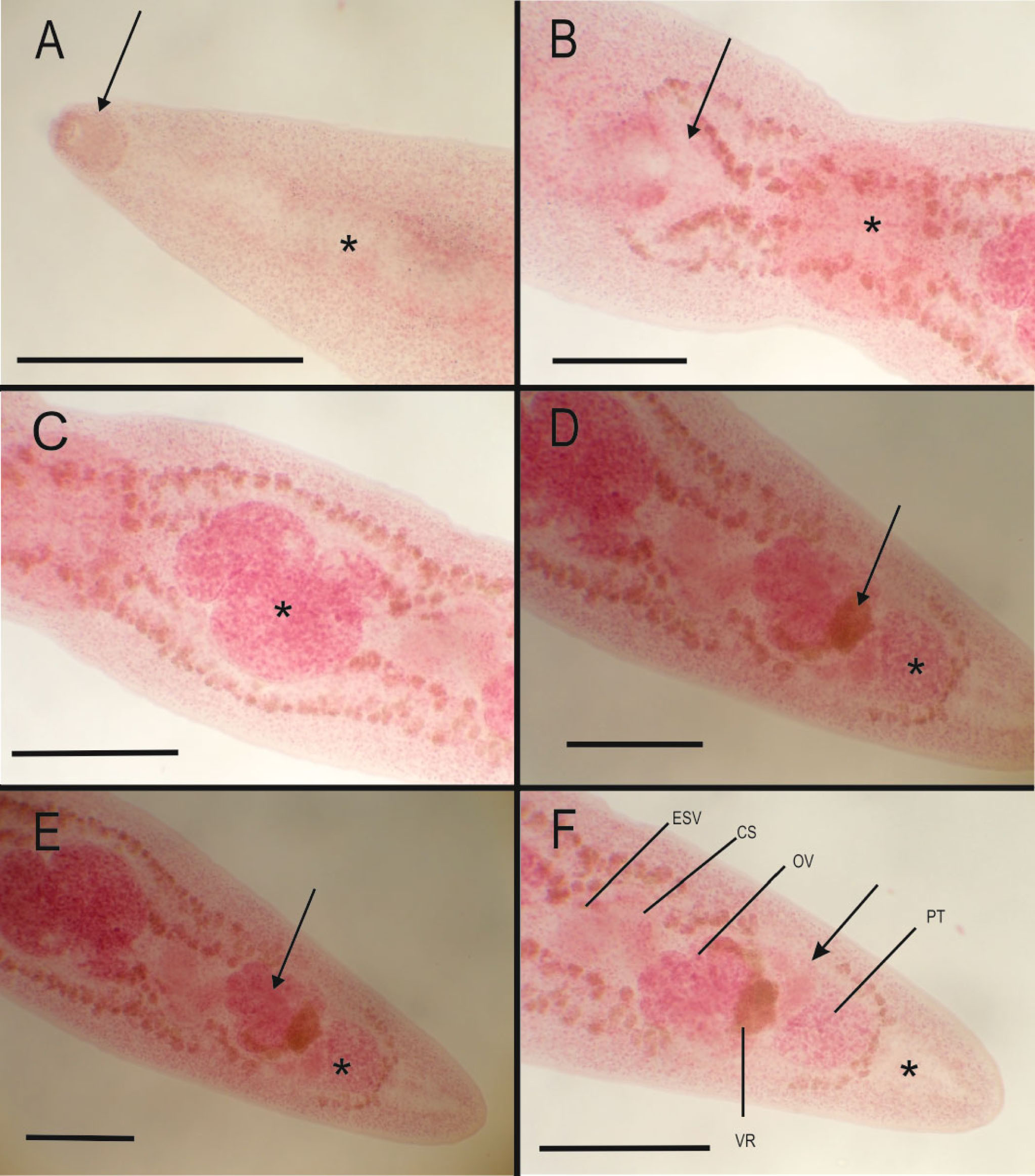
Fig. 2
Amphiorchis solus (Simha & Chattopadhyaya, 1970) Platt, 2002 (Digenea: Spirorchiidae) found in Chelonia mydas Linnaeus 1758 (Testudines, Cheloniidae) from Brazil.
(A) Oral sucker (black arrow) and esophagus (asterisk) (scale bar = 500 μm). (B) Intestinal bifucartion (black arrow) and acetabulum (asterisk) (scale bar = 250 μm). (C) Anterior testis (asterisk) (scale bar = 250 μm). (D) Dorsal view-Vitelline reservoir (black arrow) and posterior testis (asterisk) (scale bar = 200 μm). (E) Dorsal view-Ovary (black arrow) and posterior testis (asterisk) (scale bar= 200 μm). (F) Ventral view-Uterus (black arrow) and excretory vesicle (asterisk); ESV= external seminal vesicle, CS= Cirrus sac, OV= Ovary, PT= Posterior testis, VR= vitelline reservoir (scale bar = 250 μm)