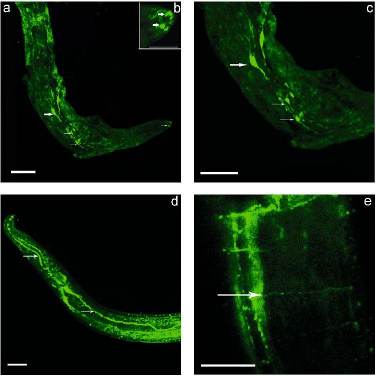
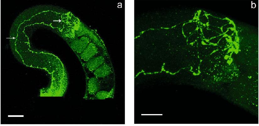
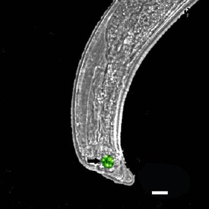
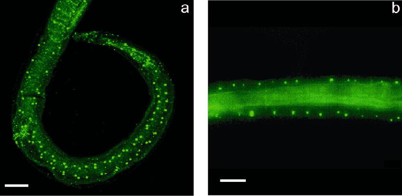
Have a personal or library account? Click to login

Serotonin in Trichinella pseudospiralis: An immunocytochemical study

Open Access
|Apr 2016

Figures & Tables

Fig. 1

The pattern of 5-HT-IR neurons and fibres in head region of I pseudospiralis. a – Three pairs of neurons (large and small arrows) with nerve fibres extending in anterior and posterior direction. Note 5-HT-IR in the tip of the head (small arrow). Bar = 30 μm. Inset. Magnification of the tip of the head with 5-HT-IR structures (arrows); b-Bar= 10 μm; c- Magnification of the three pairs of neurons (large and small arrows). Bar =25 μm; d- The pattern of 5-HT nerve cords (arrows). Bar = 20 μm; e – Magnification of the longitudinal nerve cord connected by transverse commissures (arrow). Bar =10 μm

Fig. 2

5-HT-IR nerve cells and fibres forming a plexus near of vulva in I pseudospiralis (the nerve trunks are noted by arrows also). a-Bar =20 μm; b-Large magnification of the 5-HT-IR nerve cells and fibres near of vulva Bar =10 μm

Fig. 3

5-HT-IR nerve cells in the tail of male of T. pseudospiralis. Bar =10 μm

Fig. 4

a, b – 5-HT-IR structures representing sublateral rows of hypodermal glands along the body of T. pseudospiralis. Bar =20 μm
DOI: https://doi.org/10.1515/helmin-2016-0002 | Journal eISSN: 1336-9083 | Journal ISSN: 0440-6605
Language: English
Page range: 113 - 119
Submitted on: Dec 22, 2015
Accepted on: Mar 9, 2016
Published on: Apr 22, 2016
Published by: Slovak Academy of Sciences, Mathematical Institute
In partnership with: Paradigm Publishing Services
Publication frequency: 4 issues per year

© 2016 N. B. Terenina, N. B. Mochalova, I. M. Odoevskaya, N. D. Kreshchenko, M. K. S. Gustafsson, H-P. Fagerholm, published by Slovak Academy of Sciences, Mathematical Institute
This work is licensed under the Creative Commons Attribution-NonCommercial-NoDerivatives 3.0 License.