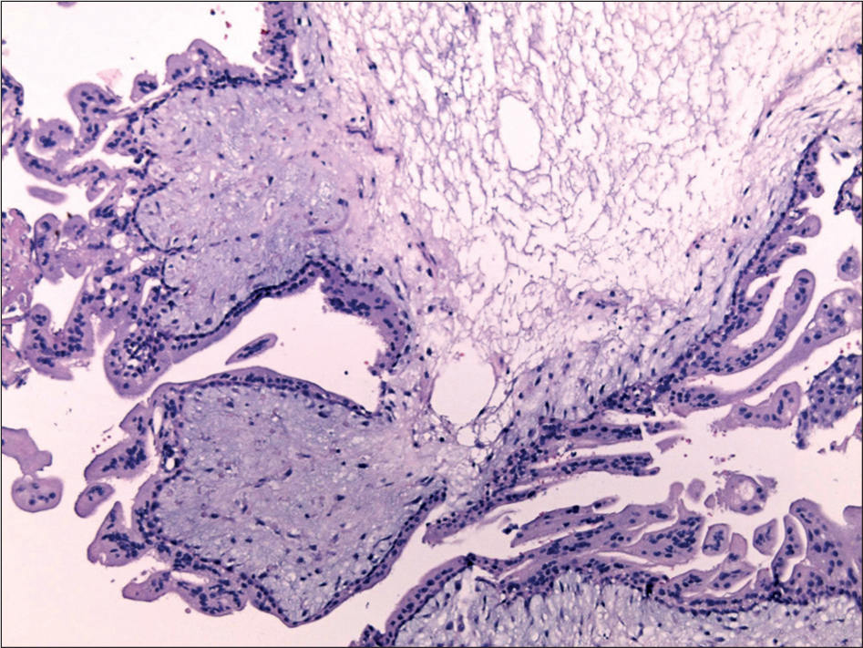
Figure 1
Microscopic appearance of the chorionic villi in complete hydatidiform mole with a pronounced stromal edema and trophoblastic proliferation (hematoxylin and eosin, ×100).
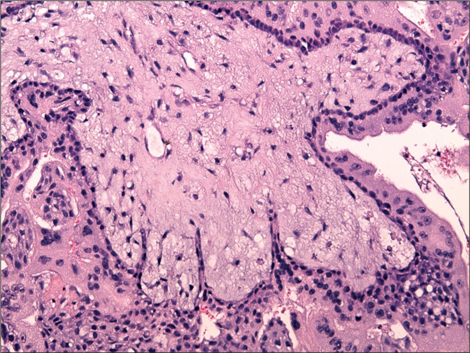
Figure 2
Mucoid stromal degeneration and irregular villous contours in complete hydatidiform mole (hematoxylin and eosin, ×200).
Table 1
Primary antibodies with clones and dilutions.
| Primary Antibody (producer) | Clone | Dilution |
|---|---|---|
| Monoclonal mouse anti-human Ki-67 antigen (Ventana) | MIB-1 | RTU |
| Monoclonal mouse primary antibody anti-p63 (Ventana) | 4A4 | RTU |
| Monoclonal mouse Primary antibody anti-p57 (Vector Laboratories) | Kip2 | 1:50 |
[i] RTU: ready to use.
Table 2
Histopathological characteristics of complete mole.
| Complete Mole Characteristics | Absent or Present | Complete Mole Cases n = 8 (%) |
|---|---|---|
| Heterogenous population of villi | absent present | 0 (0.0) 8 (100.0) |
| Circumferential trophoblastic proliferation | absent present | 1 (12.5) 7 (87.5) |
| Cytotrophoblastic proliferation | absent present | 3 (37.5) 5 (62.5) |
| Nuclear atypia | absent present | 0 (0.0) 8 (100.0) |
| Karyorrhectic stromal debris | absent present | 6 (75 0) 2 (25.0) |
Table 3
Histopathological characteristics of partial mole.
| Partial Mole Characteristics | Absent or Present | Complete Mole Cases n = 8 (%) |
|---|---|---|
| Two types of villi | absent present | 8 (100.0) 0(0.0) |
| Irregular villus contours | absent present | 0(0.0) 8 (100.0) |
| Trophoblastic stromal inclusions | absent present | 7(87.5) 1 (12.5) |
| Focal trophoblastic proliferation | absent present | 7(87.5) 1 (12.5) |
| Presence of fetal erythrocytes | absent present | 8 (100.0) 0 (0.0) |
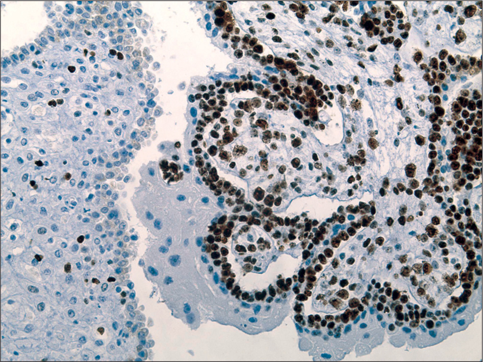
Figure 3
Ki-67 proliferation index >50.0% in villous cytotrophoblasts (×200).
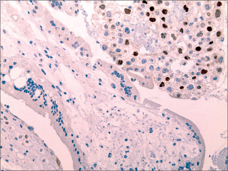
Figure 4
Absence of p57 staining In the villous cytotrophoblasts and villous stromal cells (×200).
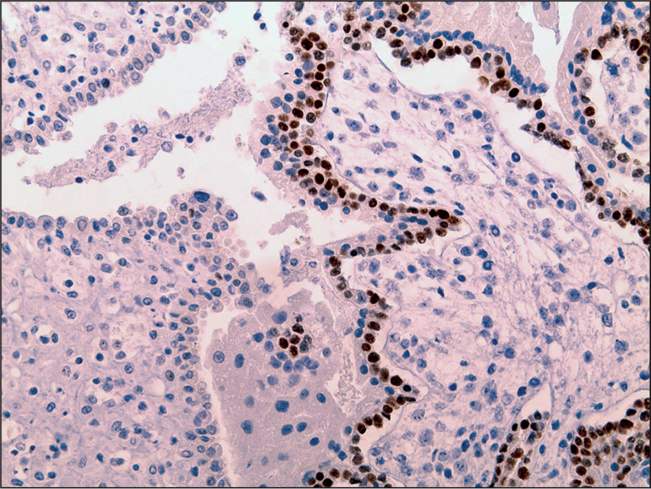
Figure 5
p63 staining in >50.0% of the villous cytotrophoblasts (×200).
Table 4
Immunohistochemical characteristics of complete moles.
| Positive Cells (%) | Ki-67 Antibody n = 8 | p63 Antibody n = 8 | p57 Antibody n = 8 |
|---|---|---|---|
| n (%) | n (%) | n (%) | |
| 0 | 0 (0.0) | 0 (0.0) | 8 (100) |
| 10 | 0 (0.0) | 0 (0.0) | 0 (0.0) |
| 20 | 0 (0.0) | 0 (0.0) | 0 (0.0) |
| 30 | 0 (0.0) | 0 (0.0) | 0 (0.0) |
| 40 | 0 (0.0) | 0 (0.0) | 0 (0.0) |
| 50 | 0 (0.0) | 0 (0.0) | 0 (0.0) |
| 60 | 0 (0.0) | 3 (37.5) | 0 (0.0) |
| 70 | 4 (50.0) | 2 (25.0) | 0 (0.0) |
| 80 | 3 (37.5) | 3 (37.5) | 0 (0.0) |
| 90 | 1 (12.5) | 0(0.0) | 0 (0.0) |
| 100 | 0 (0.0) | 0(0.0) | 0 (0.0) |