
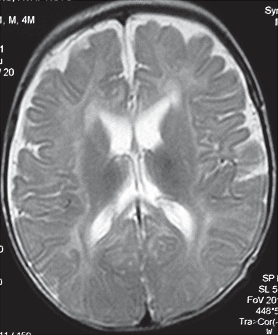
Brain MRI of the first patient at the age of 3.5 months (T2 weighted sequences TR/TE 4000/99). Note the mildly increased signal in the posterior limbs of the internal capsule bilaterally and in the white matter of the occipital lobe.
Table 1
Nerve conduction velocities in both patients.
| Nerve Conduction Velocities (m/s) | ||||||
|---|---|---|---|---|---|---|
| Age When Examined | Median Nerve | Ulnar Nerve | Tibial Nerve | Peroneal Nerve | Sural Nerve | |
| Patient 1 | 4 months | 28.2 | 25.0 | 21.4 | 18.7 | 33.2 |
| Normal values for 1-6 months | 33.3 | 35.0 | 35.0 | 38.8 | 40.6 | |
| Patient 2 | 18 months | 30.2 | 31.8 | 29.2 | 34.6 | 29.4 |
| Normal values for 1-2 years | 53.6 | 52.2 | 50.2 | 51.4 | 53.0 | |
Normal values (represent the lower normal limit [mean ± 2 standard deviation (SD)] for NCV at the age of 4 months in our laboratory.
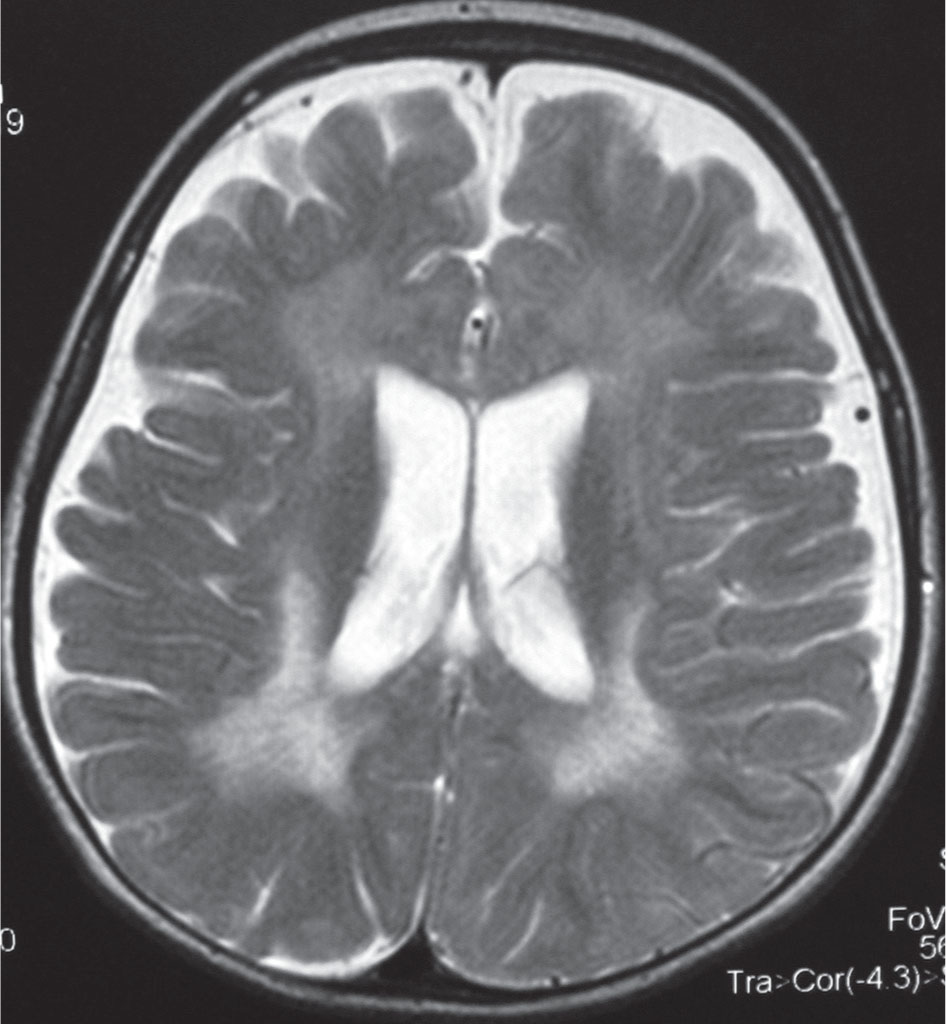
Brain MRI of second patient at the age of 18 months (T2 weighted sequences TR/TE 4510/119). Note the increased signal in the periventicular white matter around the occipital horns of the lateral ventricles with enlargement of both lateral ventricles and the subarachnoid spaces.