
A 62-year-old female with morbid obesity, hypertension, and hyperlipidemia presented to the emergency department for evaluation of atypical substernal chest pressure and shortness of breath. The symptoms had lasted 6 to 8 weeks with a crescendo pattern. An electrocardiogram on admission showed normal sinus rhythm without ischemic changes. The 0- and 3-hour cardiac enzymes (troponin I) were minimally elevated without a significant delta. Given the patient’s low GRACE score, we pursued an ischemia-guided strategy and performed a positron emission tomography (PET) scan. The patient underwent a standard rest-stress pharmacologic (regadenoson) PET with rubidium-82. The stress echocardiogram showed no ischemic changes, and the ejection fraction (EF) did not increase at peak stress (rest EF 70%, stress EF 67%). The perfusion images revealed a large, moderate to severe intensity, completely reversible perfusion defect in the basal to distal anterior and antero-septal segments that was suspicious for ischemia in the left anterior descending (LAD) coronary artery vascular territory (Figure 1A). Surprisingly, the patient’s calcium score (performed as a standard part of the PET exam) was zero (Figure 1B). This raised concern of a false positive perfusion study and prompted us to reinspect the technical aspects of image acquisition; however, no technical errors were identified. Myocardial blood flow analysis revealed abnormal flow augmentation in the LAD territory (peak stress flow in the LAD 1.3 mL/min/g; expected normal > 1.8 mL/min/g) and normal flow augmentation in the rest of the coronary vessels (Figure 1C). The PET scan was abnormal with 26% ischemia in the LAD vascular territory. The patient underwent coronary angiography, revealing a 90% proximal LAD lesion, which was treated with a (3.0 × 15) Resolute Onyx drug-eluting stent (Medtronic; Figure 1D and 1E). The remaining coronary arteries were free of atherosclerotic disease.
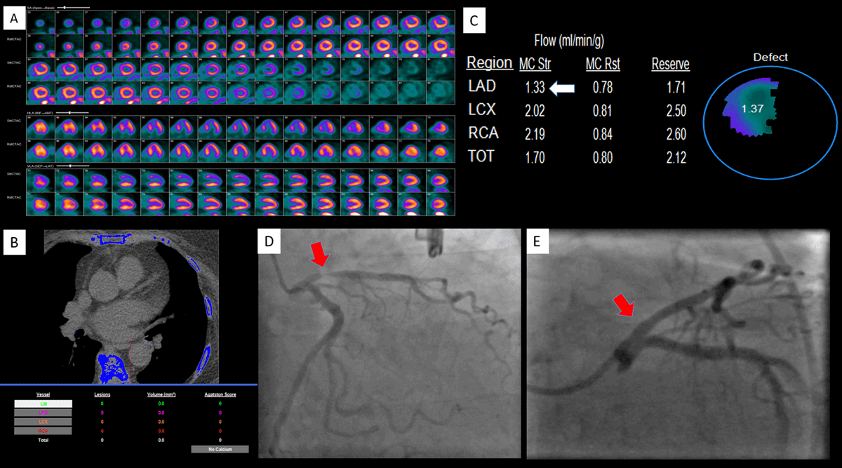
Figure 1
(A) PET perfusion images reveal a large, moderate to severe, reversible, myocardial perfusion defect in the LAD coronary artery vascular territory. (B) The calcium score was zero. (C) Myocardial blood flow analysis revealed reduced peak stress blood flow in the LAD territory but preserved global myocardial flow reserve. (D) Invasive angiographic images reveal severe stenosis in the proximal LAD that was (E) treated successfully with a drug-eluting stent. PET: positron emission tomography; LAD: left anterior descending artery.
Acknowledgement
The authors are grateful to our colleagues who provide great clinical care to our patients.
Competing Interests
The authors have no competing interests to declare.