
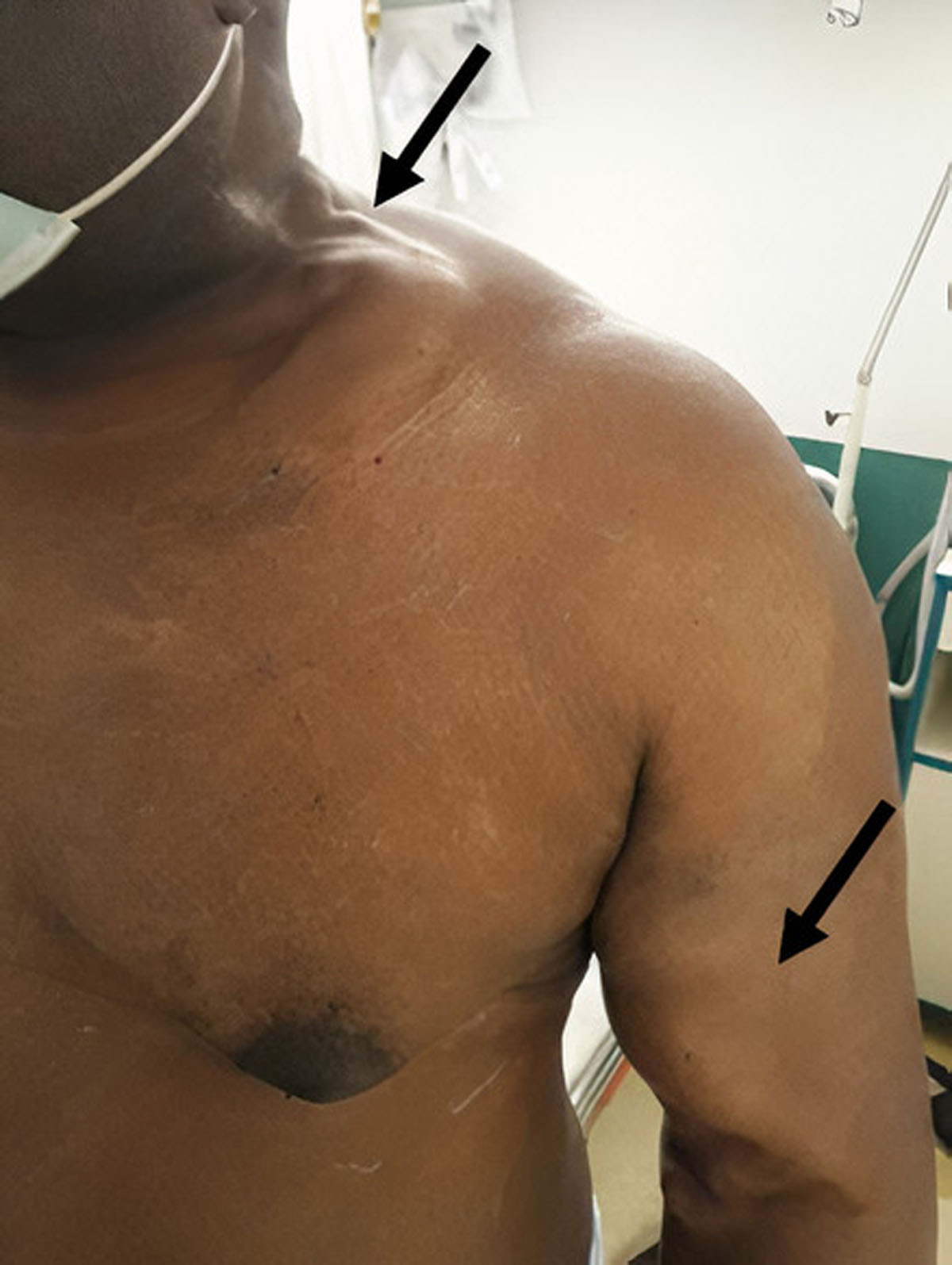
Figure 1
Aneurysmal brachiocephalic arteriovenous fistula and prominent axillosubclavian and external jugular veins in the left extremity.
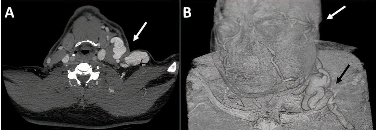
Figure 2
(A) Computed tomographic image shows retrograde dilatation of the left axillosubclavian and external jugular veins and (B) the venous drainage of the cranio-cervico-facial region.
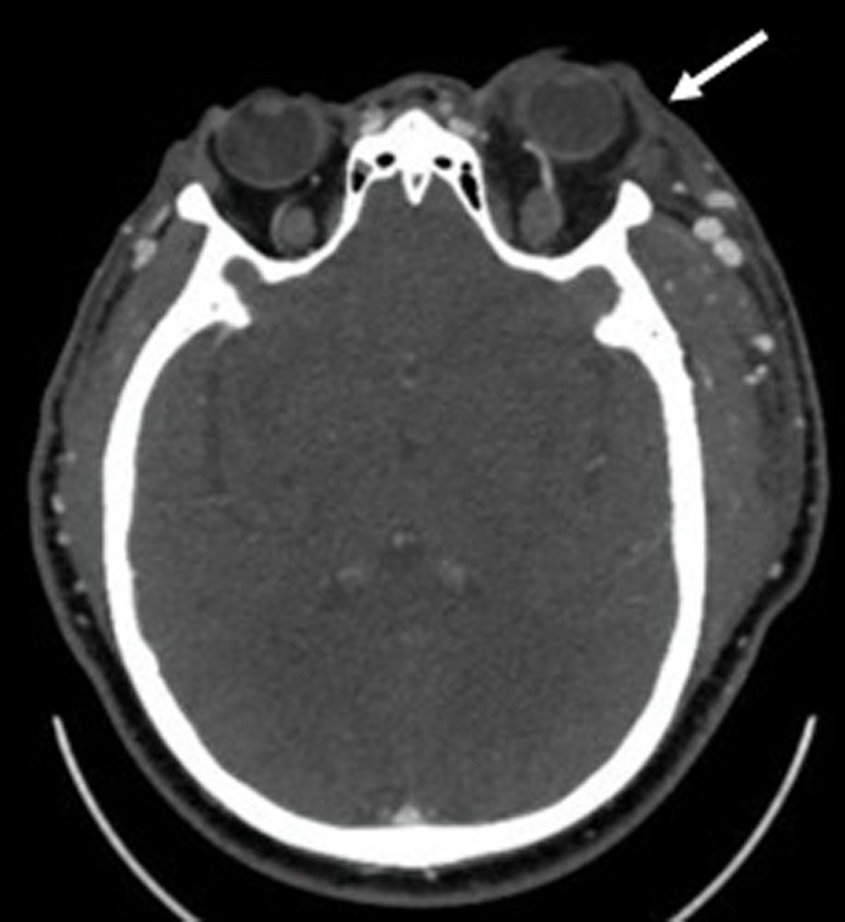
Figure 3
Computed tomographic image shows dilatation of the left superior ophthalmic vein and proptosis of the left eye in relation to an increased retrograde venous flow.