
Introduction
The association between severe elevation in blood cholesterol levels and early atherosclerotic cardiovascular disease (ASCVD) was first described more than 80 years ago by Müller, who reported families in Norway with hereditary xanthomatosis and angina.1 This report is considered the first description of familial hypercholesterolemia (FH) and included some individuals with the most severe form of the disease, homozygous FH (HoFH). The disorder was characterized initially by extreme elevations in total cholesterol, tendinous xanthomas over the extensor tendons (the metacarpophalangeal joints, knees, elbows, and especially Achilles tendons), familial clustering among first-degree relatives, and premature atherosclerosis. Consanguinity and endogamy later became recognized features and formed the basis for founder effects observed in several genetic disorders.
Although population estimates vary, large population-based meta-analyses have estimated the prevalence of heterozygous FH (HeFH) at approximately 1 in 311.2,3 In founder populations, the prevalence may be substantially higher, underscoring the importance of regional awareness, screening, and cascade detection strategies. This estimate has largely been confirmed in large-scale genome cohorts, including the 100,000 Genome Project (1/288)4 and the All of Us cohort (1/287).5 Based on a United States (US) population of more than 342 million, these estimates suggest that there are well over 1 million FH cases in the US.
Here, we review the definition and genetic basis of FH, the burden of disease, particularly ASCVD and aortic stenosis, the importance of registries, and current therapeutic modalities. We also address early identification in youth and sex differences in diagnosis and treatment. Finally, we discuss the most severe form of FH, HoFH.
Interestingly, FH and its treatment with statins represent one of the great unsung successes of medicine. The natural history of untreated FH leads to premature ASCVD morbidity and mortality, whereas prompt recognition and treatment can lead to a normal life expectancy.6
Definition and Genetic Diagnosis
Clinical Diagnostic Criteria
The phenotype of FH has changed over the past several decades, with definitions varying across countries of origin and professional associations. Original definitions include the Dutch Lipid Clinic Network and the Simon Broome criteria, which are still widely used.6,7 However, these definitions emphasize the presence of tendinous xanthomas as well as arcus senilis and xanthelasmas, the latter two being much less specific for FH. These clinical signs are now seen less often because of earlier diagnosis, greater reliance on biochemical parameters, particularly LDL-C levels, and early statin therapy. Ahmad et al.7 recently reviewed this topic for the US National Lipid Association. The major diagnostic criteria include absolute LDL-C levels, adjusted for age, affected first-degree relatives, premature ASCVD, and tendinous xanthomas. A simplified Canadian definition for FH has also been developed to facilitate clinical diagnosis and harmonize case identification across lipid clinics.8
Although once considered the “gold standard,” identifying a pathogenic variant in an FH-related gene is not essential for clinical diagnosis. Genetic testing is increasingly accessible in the United States for patients with insurance coverage, but some limitations remain. First, a proportion of patients who meet clinical criteria for FH do not have a detectable pathogenic variant with current next-generation sequencing methods. Second, while molecular confirmation can increase diagnostic certainty, refine risk stratification, and facilitate cascade screening, treatment decisions are generally guided by LDL-C levels, clinical risk, and family history.
Currently, the major diagnostic criterion is an elevated plasma level of LDL-C, usually > 95th percentile for age and sex. In the US, this corresponds to 190 mg/dL (5.0 mmol/L) for men and women older than 40 years. Some medical conditions can be associated with marked elevations in LDL-C (Table 1). Importantly, screening for these conditions should be performed as part of the initial evaluation.
Table 1
Selected conditions associated with marked low-density lipoprotein cholesterol elevation. TSH: thyroid-stimulating hormone
| CONDITION | SCREENING |
|---|---|
| Hypothyroidism | TSH, T4, T3 |
| Nephrotic syndrome | Urinary protein |
| Biliary cirrhosis | Liver function tests |
| Ketogenic diet | History |
Rare genetic disorders
| Specialized genetic testing (referral) ABCG5/8 gene complex CYP27A1 LIPA ALB |
An algorithm for the diagnosis of FH, adapted from the simplified Canadian definition, is proposed in Figure 1. A diagnosis of “definite” FH is based on the cumulative findings of very high LDL-C levels, adjusted for age, a family history of affected first-degree relatives with elevated LDL-C, premature ASCVD, tendinous xanthomas, and a genetic diagnosis. “Probable” FH includes LDL-C levels and family history, whereas “possible” FH may be based solely on an elevated LDL-C level. Often, information on family members is unavailable and xanthomas may not have developed because of younger age or current treatment with statins. This group of patients poses a challenge for clinicians.
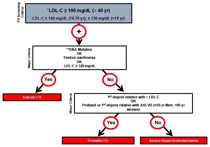
Figure 1
Simplified algorithm for the diagnosis of familial hypercholesterolemia (FH). Modified from Ruel 2018 and Ahmad 2026.7
With statin therapy being ubiquitous in the US population, a baseline untreated LDL-C value may not be available, even in electronic medical records. Ruel et al.8 provided a means to impute the untreated LDL-C in FH patients from the current on-treatment value and the type and dose of statin and ezetimibe used. A web-based calculator is available at https://www.circl.ubc.ca/english/web_ldl2.html.
The new International Classification of Diseases, 10th Revision (ICD-10) codes in the United States include E78.011 for heterozygous FH, E78.010 for HoFH and E78.019 for “unspecified” FH. In patients who meet the LDL-C criterion but lack information on family history and do not have xanthomas, a molecular confirmation may be required to establish a “definite FH” diagnosis, encourage family cascade screening, and provide access to medications such as proprotein convertase subtilisin/kexin type 9 (PCSK9) inhibitors.
Molecular Basis of FH
The molecular basis for FH was elucidated by Drs. Goldstein and Brown and led to the Nobel prize for the discovery of receptor-mediated endocytosis and the LDL receptor (LDL-R).9 LDL particles contain one molecule of apolipoprotein B (ApoB), which binds to the LDL-R. Another protein, PCSK9, modulates cell-surface expression of the LDL-R. Pathogenic variants in the genes (LDLR, APOB, and PCSK9) coding for these proteins can cause FH. Variants in the LDL-R adaptor protein 1 (LDLRAP1) cause an autosomal recessive form of the disease (Figure 2).10
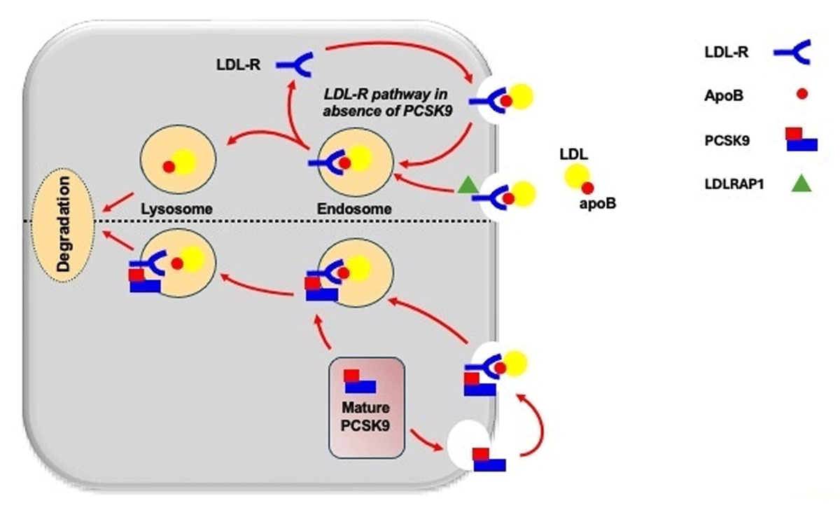
Figure 2
LDL receptor-mediated uptake of low-density lipoprotein (LDL) particles and PCSK9-mediated LDL receptor degradation. Pathogenic variants in LDLR, APOB, PCSK9, and LDLRAP1 cause familial hypercholesterolemia. Modified with permission from Awan et al.
It should be noted that pathogenic variants in PCSK9 causing FH are gain-of-function variants. These are seen much less frequently than variants in LDLR or APOB genes. However, the discovery of gain-of-function variants in PCSK9 provided a therapeutic avenue for the development of PCSK9 inhibitors for the treatment of severe hypercholesterolemia.
Genotype-Phenotype Considerations
Furthermore, pathogenic variants in the LDLR gene alter the function of the protein across a spectrum ranging from reduced receptor activity (“defective” variants) to complete absence of LDL-R activity on the cell surface (“null” variants). These correlate with LDL-C levels, age of onset, and severity of cardiovascular disease. Some individuals with a documented pathogenic variant at the LDLR or APOB genes may have an LDL-C below the threshold for FH because of an excellent lifestyle or the presence of another genetic variant associated with lower LDL-C levels.
In addition, the cumulative effect of small variants in several genes that regulate LDL-C levels in an individual, referred to as polygenic hypercholesterolemia, may present with LDL-C levels in the range of FH, but the pattern of inheritance is not classically Mendelian.11 As many as 30% to 40% of patients with FH diagnosed on clinical grounds have a polygenic form of hypercholesterolemia.
Despite advances in DNA sequencing technologies, some patients with a clinical diagnosis of FH may have variants deeply buried in introns of known genes or in a novel gene not yet identified in FH. The genes associated with FH are shown in Table 2.
Table 2
Genetic basis for FH (modified from Ahmad 20267). LDLR: low-density lipoprotein receptor; FH: familial hypercholesterolemia; LDL-C: low-density lipoprotein cholesterol
| GENE | MODE OF INHERITANCE | PERCENT OF FH CASES | |
|---|---|---|---|
| LDLR | Autosomal semi-dominant | ~85%–90% | Over 2,000 variants identified. Reduce LDLR function (null or defective alleles). Clinical severity varies by mutation type |
| APOB | Autosomal semi-dominant | ~5%–10% | Impair binding of LDL particles to LDLR. Milder phenotype than LDLR mutations |
| PCSK9 | Autosomal semi-dominant | 1%–3% | Gain-of-function mutations increase LDLR degradation |
| LDLRAP1 | Autosomal recessive | Rare | LDLR endocytosis is defective |
| Polygenic hypercholesterolemia | Cumulative effect of multiple genes causing small elevations in LDL-C | ~30–40% of clinically diagnosed FH | Non-Mendelian inheritance |
The term heterozygous FH (HeFH) is used to describe patients who meet diagnostic criteria and have a single pathogenic variant. Homozygous FH (HoFH) is now described as bi-allelic semidominant monogenic (LDLR gene) and bi-allelic semidominant digenic (LDLR and APOB genes). A third form, the rare bi-allelic recessive hypercholesterolemia, is caused by variants in LDLRAP1 inherited from each parent.
The LDLR gene underlies most FH cases. Well over 2,000 pathogenic variants have been reported, and in-depth analyses of most DNA coding variants have revealed the wide range of physiological impact of individual mutations.12
Cardiovascular Risk, Atherosclerosis, and Aortic Stenosis
Since its original description, FH has been associated with premature ASCVD. The advent of statins and other lipid-lowering therapies has dramatically altered the natural course of the disease, and patients with HeFH who are identified and treated early can expect a normal life expectancy.6 Current cholesterol guidelines consider FH as an ASCVD equivalent and recommend early detection and treatment.6,13,14
The risk of ASCVD is related to the lifelong burden of LDL-C. In a study of 21,426 individuals without coronary disease from six US cohort studies—including the Atherosclerosis Risk in Communities study, Coronary Artery Risk Development in Young Adults study, Cardiovascular Health Study, Framingham Heart Study Offspring cohort, Jackson Heart Study, and Multi-Ethnic Study of Atherosclerosis—the presence of an FH variant was associated with increased cardiovascular risk, with a hazard ratio of 2.9 (95% CI, 1.4-6.0).15 In 245,388 participants from the All of Us project, the presence of a pathogenic FH variant increased cardiovascular risk by 2.91-fold (95% CI, 2.34-3.58).5 These contemporary studies reflect the US population and may not be generalizable to other countries, especially those with lower socioeconomic status or different access to lipid-lowering therapies. Cardiovascular disease is strongly related to LDL-C levels; Figure 3 displays the continuum of ASCVD risk across plasma LDL-C levels and genetically confirmed FH. The burden of lifelong LDL-C elevation can be roughly determined using the cholesterol-year score.
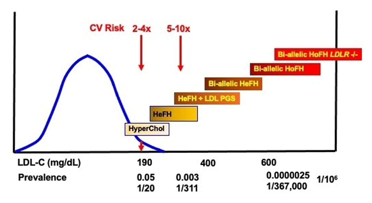
Figure 3
Continuum of cardiovascular risk across LDL-C levels. A population distribution curve for LDL-C in adults is shown in blue, with the 95th percentile at 190 mg/dL. Increasing severity of the FH phenotype increases ASCVD risk. LDL-C: low-density lipoprotein cholesterol; ASCVD: atherosclerotic cardiovascular disease; FH: familial hypercholesterolemia; HeFH: heterozygous FH; HoFH: homozygous FH; HyperChol: hypercholesterolemia; LDL PGS: low-density lipoprotein polygenic score
Screening for ASCVD in patients with FH is widely recommended. Most current guidelines exclude FH from standard cardiovascular risk algorithms and suggest that markedly elevated LDL-C constitutes an indication for statin therapy.13,14 Specific risk scores for ASCVD in patients with FH, including Canadian and French models, have been developed and, unsurprisingly, include conventional cardiovascular risk factors such as age, sex, smoking, high blood pressure, and diabetes.16 The importance of lifestyle and obesity to cardiovascular risk has also been emphasized in large, international registries.17
Noninvasive imaging techniques help refine cardiovascular risk stratification in patients with FH and support treatment intensification, particularly when there is uncertainty about risk, evidence of additional risk modifiers, or concern for subclinical atherosclerosis. These techniques include carotid ultrasonography to detect the presence of atherosclerotic plaques and coronary calcium scoring by computed tomography (CT). In younger patients, atherosclerotic plaques can be present without calcification, limiting the sensitivity of coronary calcium scoring. In selected cases, coronary CT angiography (CCTA) can provide additional information on non-calcified plaque burden and coronary anatomy.
The optimal timing, frequency, and choice of imaging modality in asymptomatic patients with FH remain incompletely defined and should be individualized based on age, cumulative LDL-C burden, family history, symptoms, and other risk modifiers. CCTA provides anatomical resolution approaching that of conventional angiography. However, the use of contrast media, radiation exposure, and uncertain clinical value in some asymptomatic patients are important considerations. Nevertheless, the presence of subclinical or clinical ASCVD in a patient with FH supports intensive LDL-C lowering, often beyond statins and ezetimibe, in accordance with current guidelines.13,14
The development of aortic stenosis increases dramatically with age in the general population.18,19,20 Mendelian randomization studies show that variants at the LDLR gene locus are associated with aortic stenosis.21,22 A recent meta-analysis of aortic stenosis in FH reveals that patients with HeFH are at a fourfold increased risk of aortic stenosis compared to the general population, as well as an increased need for aortic valve replacement.23
Registries and Prognosis
Many countries have established registries for FH to raise awareness, collect real-world data and inform regulators on the burden of disease and the need for screening, diagnosis, and treatment.24 In the US, the CASCADE FH registry is an ongoing project,25 closely affiliated with the Family Heart Foundation (https://www.familyheart.org/), which has led efforts to raise awareness of familial hypercholesterolemia and elevated lipoprotein(a) while fostering collaborative research and knowledge dissemination. The European Atherosclerosis Society (EAS) supports the Familial Hypercholesterolemia Studies Collaboration (https://eas-fhsc.org/), a worldwide initiative that initially collected data on FH from 56 countries and has grown to include more than 70 countries.26
Registries also provide an important platform for cascade screening, longitudinal follow-up, quality improvement, and evaluation of treatment gaps. Overall, country-specific registries reveal that FH remains underdiagnosed and undertreated, with persistent sex differences in diagnosis and treatment. In the US, despite an increase in specialized lipid clinics and available treatments, elevations in LDL-C > 100 mg/dL were found in 52% of patients, prompting calls for earlier and more intensive treatment of FH. The worldwide discrepancies in outcomes between high- and low-income countries were also brought to light with the Familial Hypercholesterolemia Studies Collaboration publications.27 In the US, several challenges remain including case identification, cascade screening, access to expensive medications, and insurance coverage.
Treatment
The treatment of FH must involve a careful evaluation of lifestyle and diet. Because the risk of ASCVD in patients with FH is much higher than age- and sex-matched individuals, a healthy lifestyle should be encouraged and supported. This includes the quality of diet, attaining a healthy body weight, regular physical activity and smoking cessation. Despite these measures, patients rarely achieve guideline-recommended therapeutic goals with lifestyle modification alone.25
Initial pharmacologic treatment should consist of a high-potency statin, such as rosuvastatin 20 mg to 40 mg or atorvastatin 40 mg to 80 mg daily. If side effects limit adherence to treatment, reducing the dose of statin and adding ezetimibe 10 mg can produce up to 50% LDL-C reduction. Several algorithms for the treatment of FH have been proposed.7 In cases where the combination of statin and ezetimibe is insufficient, PCSK9 inhibitors have proven remarkably effective in further lowering LDL-C. These include the monoclonal antibodies evolocumab (Repatha®) and alirocumab (Praluent®).27 The siRNA inclisiran (Leqvio®) directed at PCSK9 messenger RNA has also been approved for the treatment of FH.
In statin-intolerant patients, the liver-specific adenosine triphosphate (ATP) citrate lyase inhibitor bempedoic acid (Nexletol®) provides an alternative or additional oral therapy to statins.28 The cholesteryl ester transfer protein inhibitor obicetrapib may also become an additional oral option for patients with FH.29 The development of macrocyclic peptides as absorbable peptide-like medications may represent an important development in pharmacotherapy. Enlicitide (Lipfendra®), the first approved oral PCSK9 inhibitor, has shown similar LDL-C lowering efficacy, approaching that of injectable monoclonal antibodies.30
Most FH experts recommend initiating statin therapy in children with FH from 8 to 10 years of age. This decision should consider the presence of ASCVD in parents or affected siblings and should occur with close follow-up in an experienced center.13,14
Women and Pregnancy
Registries have highlighted important sex-based differences in the recognition and management of FH, showing that women are often diagnosed later and receive less intensive treatment than men.31,32 Yet, ASCVD events generally occur later in women, most likely because of the relative cardiovascular protection associated with the premenopausal years.27 Pregnancy and lactation represent important challenges in the care of women with FH. Statins and most other cholesterol-lowering drugs are generally discontinued during pregnancy and lactation, although inadvertent statin exposure in early pregnancy has not been associated with a clear increase in congenital malformations in observational studies.33 Women with FH during their reproductive years may therefore face prolonged periods without lipid-lowering therapy, particularly during pregnancy, breastfeeding, and attempts to conceive.34,35 Management requires preconception counseling, individualized risk assessment, and coordination between lipid specialists, obstetricians, and primary care providers.
Homozygous Familial Hypercholesterolemia
Based on an estimated worldwide prevalence of HeFH at 1 in 311 individuals, the expected prevalence of HoFH is approximately 1 in 386,000, which would predict approximately 1,000 cases in the US. The widely accepted definition of HoFH follows the EAS position statement on HoFH.36 An untreated elevated LDL-C above 10 mmol/L or 400 mg/dL (the 95th percentile for individuals < 20 years of age is ~130 mg/dL) is suggestive of HoFH and requires further evaluation, including detailed clinical and family history and genetic testing when available. Additional clinical features include cutaneous or tendon xanthomas before 10 years of age and/or untreated LDL-C levels consistent with HeFH in both parents, although in digenic forms one parent may have normal LDL-C levels and the other may have LDL-C levels consistent with HoFH.
Since the identification of genes involved in FH, the definition of HoFH has been extended to reflect its genetic complexity. The 2023 EAS statement emphasizes that HoFH is best understood as an umbrella term encompassing several genetic diagnoses. Genetic confirmation includes biallelic pathogenic or likely pathogenic variants on different chromosomes in LDLR, APOB, PCSK9, or LDLRAP1, or pathogenic variants at different FH-associated loci. These include biallelic semi-dominant monogenic hypercholesterolemia caused by two identical or two different variants in the same gene; biallelic semi-dominant digenic hypercholesterolemia, caused by variants in two different FH-associated genes; and the rare biallelic recessive form caused by LDLRAP1 variants.
The most severe form of HoFH is generally associated with biallelic LDLR “null” variants,7,36 which result in absent or near-absent LDL receptor function and are associated with a more severe clinical course and reduced responsiveness to LDL receptor-dependent therapies than “defective” variants, which retain some LDLR function. However, genotype does not always predict the precise phenotype in an individual patient, and in cases of discordance, the phenotypic diagnosis should take priority over the genetic result.
Patients with HoFH usually have normal physical, cognitive, and sexual development but may experience a marked reduction in their quality of life because of frequent medical visits, hospital admissions for cardiovascular disease, and, in some patients, extracorporeal LDL filtration through plasmapheresis or LDL apheresis.
Untreated HoFH patients rarely survive beyond the third decade of life.37,38 Over the past three decades, survival has more than doubled, especially in high-income countries.39,40 Severe, premature ASCVD often manifests in youth and young adulthood.41,42 Aortic valve disease is an important complication of HoFH and its phenotype has shifted from supravalvular stenosis to calcific aortic stenosis and, often, calcified ascending aorta (“porcelain aorta”).43
The diagnosis of HoFH in the United States is often made on the basis of cutaneous manifestations developing in childhood, markedly elevated LDL-C levels from birth, and, if untreated, very early ASCVD, such as myocardial infarction by age 20. Most patients are referred to a physician specializing in lipoprotein disorders. Treatment is aimed at lowering LDL-C levels as early and as intensively as possible. Survival of HoFH patients has improved substantially and doubled since the introduction of statins.43,44 The standard of care includes maximally tolerated statins and ezetimibe, an inhibitor of intestinal cholesterol absorption. Bile acid binding resins such as cholestyramine, colestipol, or colesevelam may also be used, although with very limited efficacy and tolerability that can be challenging. PCSK9 inhibitors are additionally used, as most patients cannot reach recommended treatment guidelines on currently available medications, although their efficacy depends on residual LDL receptor function and is therefore limited in patients with biallelic LDLR null variants.
The aim of therapy is to lower LDL-C levels according to current guidelines.13,36 Recommended LDL-C targets are rarely met, and treatment intensification usually requires multiple medications. Some patients require extracorporeal LDL filtration or LDL apheresis, in which LDL particles are selectively removed from the blood. This technique shares similarities with kidney dialysis and, despite being considered standard of care for severe HoFH, is not widely available.
Two “orphan” drugs are available for HoFH. Lomitapide (Juxtapid ® 5-60 mg daily) inhibits microsomal triglyceride transfer protein, thereby reducing hepatic secretion of VLDL and LDL particles.45 It requires careful dose titration and dietitian support to markedly decrease dietary fats. Gastrointestinal side effects and hepatic fat accumulation may limit its optimal use. Evinacumab (Evkeeza®) is a monoclonal antibody directed at the protein ANGPTL3 and lowers LDL-C through an LDL receptor-independent mechanism.46 The drug is given intravenously at a dose of 15 mg/kg body weight every 4 weeks. It has proven to be well tolerated and can lower LDL-C by approximately 50% on top of other lipid-lowering therapies. Patients with HoFH should be followed in centers with expertise in complex atherosclerotic cardiovascular disease and calcific aortic stenosis.47 Liver transplantation has been used in the past for HoFH, although this procedure is fraught with long-term complications related to immunosuppression and current pharmacologic options are preferable.48 Gene therapy and genome editing for HoFH are at an early experimental stage but may offer exciting therapeutic avenues in the future.49
Conclusions
Familial hypercholesterolemia is the most frequent monogenic disorder encountered in clinical medicine, with a population prevalence of ~1/311 individuals. Although increasingly recognized, FH remains underdiagnosed and undertreated. Yet, the early diagnosis and treatment of FH, especially with statins represents a major, but unheralded success of medicine. Untreated, FH leads to premature ASCVD, usually before the sixth decade of life. When diagnosed and treated early, it provides the patient with a normal life expectancy. The aim of treatment follows that of established guidelines. In addition to statins, ezetimibe, and bempedoic acid offer orally administered options. Inhibitors of PCSK9 include monoclonal antibodies and small interfering RNA therapeutics and are administered subcutaneously. Enlicitide, the first oral PCSK9 inhibitor, now offers an orally administered option for adults with hypercholesterolemia, including HeFH. The most severe form, homozygous FH, is caused by biallelic pathogenic variants from both parents and requires early diagnosis, specialized care, intensive LDL-C lowering, and, in some patients, LDL apheresis or orphan therapies.
Key Points
Familial hypercholesterolemia (FH) is a semidominant autosomal genetic lipoprotein disorder with a prevalence of approximately 1 in 311 individuals.
Diagnosis of FH is based on low-density lipoprotein-cholesterol (LDL-C) levels, affected first-degree relatives, tendinous xanthomas, premature atherosclerotic cardiovascular disease (ASCVD), and genetic testing when available.
FH is most commonly caused by genetic variants that affect LDL receptor-mediated clearance to varying degrees, from mild (“defective”) to severe (“null”).
Approximately 30% of clinical FH cases have polygenic hypercholesterolemia.
Untreated, FH leads to premature ASCVD, whereas prompt recognition and treatment can lead to a normal life expectancy.
The homozygous form is an orphan disease with a prevalence of approximately 1 in 367,000 and requires early and intensive treatments, including LDL apheresis and specialized therapies in some patients.